Bilder einer akzessorischen Brustwarze

supernumnipple__protectwyjqcm90zwn0il0_focusfillwzi5ncwymjisinkildm2xq-2736639-7751844-jpg-7553342

Inhaltsverzeichnis

Zugriff auf die Bildbibliothek

Illustrative Gallery: Clinical Cases of Supernumerary Nipple

Visual Manifestations of Supernumerary and Accessory Nipple

Visual example of a supernumerary nipple, illustrating a congenital anomaly of skin development.

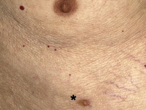
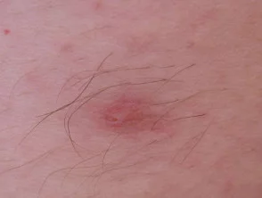
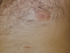
Supernumerary Nipple (Case 1)

Clinical manifestation of an accessory nipple located along the embryonic mammary ridge.

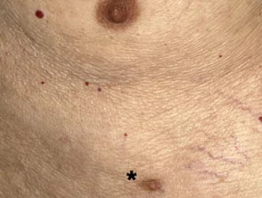
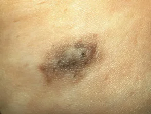
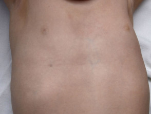
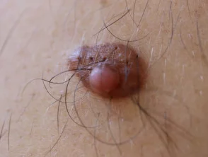
Supernumerary Nipple (Case 2)

Dermatological image detailing the characteristic morphology of a supernumerary nipple.

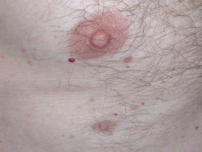
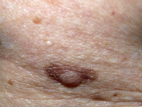
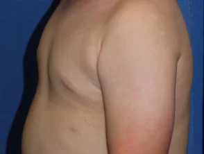
Supernumerary Nipple (Case 3)

Example of an accessory nipple clearly positioned inferiorly relative to the main breast.

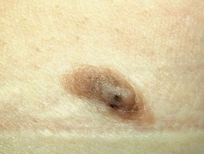
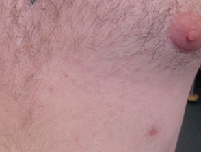
Supernumerary Nipple (Case 4)

Variant of polymastia or accessory nipple presenting a bifurcated appearance.

Supernumerary Nipple (Case 5)

Lateral view showing a supernumerary nipple with a minimal degree of tissue development.

Supernumerary Nipple (Case 6)

Illustrative image of multiple supernumerary nipples distributed along the embryonic mammary line.

Supernumerary Nipple (Case 7)

Case of a supernumerary nipple that, due to its pigmentation, could be visually confused with a common dermal nevus.

Supernumerary Nipple (Case 8)

Thorough clinical examination of an accessory nipple, highlighting its specific morphological characteristics.

Supernumerary Nipple (Case 9)

Fully developed supernumerary nipple, presenting a structure similar to a functional areola and nipple.

Supernumerary Nipple (Case 10)


Supernumerary Nipple (Case 11)

This collection of clinical images offers a detailed view of the morphological variations presented by the supernumerary nipple, a dermatological condition resulting from the ectopia of mammary tissue along the embryonic mammary line. Studying these cases helps differentiate this condition from other skin lesions.

Clinical comparison between a normal nipple and an accessory supernumerary nipple.
Supernumerary Nipple (Case 11)

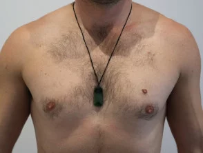
These images illustrate the morphological diversity that the supernumerary nipple can present, a congenital condition originating from the incomplete development of the fetal mammary ridges. Understanding these visual variations facilitates differential diagnosis in dermatological practice.

Macroscopic image of a supernumerary nipple
Supernumerary Nipple

Dermoscopic Analysis of the Supernumerary Nipple

Dermoscopic view of a supernumerary nipple showing characteristic vascular patterns
Dermoscopy of Supernumerary Nipple
Detailed dermoscopic examination of an accessory nipple anomaly
Dermoscopy of Supernumerary Nipple
Dermoscopic image focused on the areola of an accessory nipple
Dermoscopy of Supernumerary Nipple

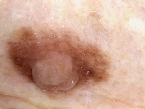
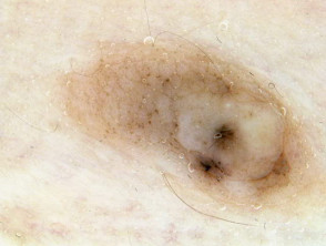
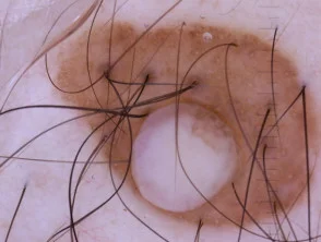
Analyzing these visual representations is crucial for the differential diagnosis of skin lesions in this area. Correct identification, facilitated by dermoscopy, confirms the presence of a supernumerary nipple by revealing specific histological characteristics under magnification.

Dermatly.com - Die Seite für Ihre Haut