Invoering
Brunsting-Perry cicatricial pemfigoïd is een zeldzame vorm van gelegen cicatriciale pemfigoïd, die vaak voorkomt in het hoofd-halsgebied. Interessant is dat het meestal niet gaat om de slijmvlies membranen zoals te zien is bij typische cicatriciale pemfigoïd. Het klinische verschil is gelokaliseerd bulleus pemfigoïd, waarbij er nauwelijks littekens zijn in vergelijking met de cicatriciale pemfigoïd van Brunsting Perry.
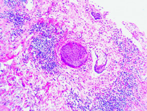
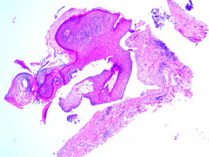
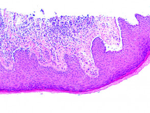
histologie Brunsting-Perry cicatriciale pemfigoïd
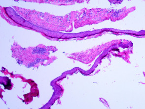
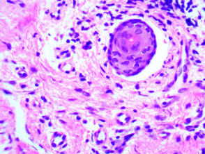
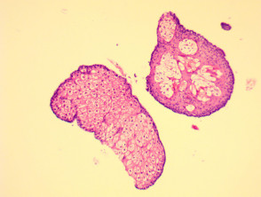
Microscopie onthult subepidermaal ampullen met verschillende mengsels van opruiend cel infiltreren. Vroeg verwondingen kan kleine papillaire tonen microabcessen
Pathologie van Brunsting-Perry cicatriciale pemfigoïd
Figuur 1
Figuur 2
figuur 3
Figuur 4
Figuur 5
Figuur 6
Afbeeldingen geleverd door Dr. Duncan Lamont, Waikato Hospital
Speciale studies over cicatriciale pemfigoïd van Brunsting-Perry
immunofluorescentie steekproef basaal membraan zone IgG en/of C3.
elektronen microscoop onthult de splitsing in de dichte sublamina met geconserveerde basaal blad en verankeren fibrillen op het dak van de ampulla.
differentiële diagnose Brunsting-Perry cicatriciale pemfigoïd
Gelokaliseerde bulleuze pemfigoïd