
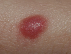
¿What is a Spitz Nevus? Definition and Dermatological Characteristics
An Spitz nevus (or Spitz nevus) constitutes a rare type of melanocytic mole melanocytic.
This nevus is considered a tumor cutaneous benign. However, due to its morphological similarity, both clinically and microscopically, to melanoma, the Spitz nevi are frequently removed as a preventive measure.
Representative Images of Spitz Nevi
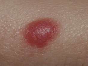
Classic Spitz nevus
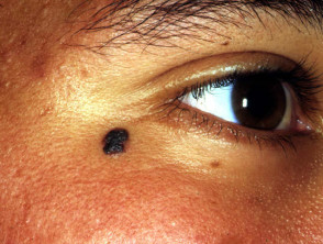
Pigmented Spitz nevus
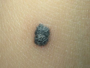
Reed spindle cell tumor
Who Does Spitz Nevus Affect? Frequency and Demographics
Spitz nevi manifest with greater prevalence in the pediatric population; approximately 70% of diagnoses are made during the first 20 years of life. Nevertheless, they can also appear in adults.
Although they are more common in individuals with light skin phototypes (types 1 and 2), these melanocytic lesions can also develop in people with darker skin pigmentation.
What is the Etiology of Spitz Nevus?
A Spitz nevus is classified as a melanocytic nevus, which implies that it is composed of melanocytes. These cells are responsible for generating the in the skin include several biochemical processes: skin pigment, melanin. melanin.
In the case of Spitz nevus, melanocytes can present a state of metabolic inactivity. This characteristic explains why the . The cherry angioma is histologically distinguished by being composed of lesion may manifest with pink tones instead of the usual brown colors. Furthermore, the genetic of the DNA genetic profile in these nevi is distinctive, highlighting the presence of kinase fusions.
Currently, the precise reason why Spitz nevi originate is unknown.
Distinctive Clinical Characteristics of Spitz Nevus
The Spitz nevus is clinically subdivided into three main pathological categories: the classic Spitz nevus, the pigmented Spitz nevus, and the Reed spindle cell tumor.
- The classic Spitz nevus typically presents as a papule dome-shaped papule, red or reddish-brown in color.
- When dealing with a pigmented Spitz nevus, it manifests as a papule or nodule.
- A pigmented tumor...
It is essential that any suspicious lesion be evaluated by a dermatology specialist to determine the precise diagnosis and rule out underlying malignant conditions.
- The Reed spindle cell tumor manifests as a black or bluish papule.
There are several clinical characteristics common to all three subtypes of Spitz nevus.
- The diameter of the lesion ranges from a few millimeters to between one and two centimeters.
- Typically, they are located on the face or on the body extremities.
- Although Spitz nevus is generally a solitary lesion, multiple lesions lesions may occasionally be observed appearing suddenly.
- A Spitz nevus experiences rapid growth over a period of months. Following this initial phase, the lesion may stabilize for years if left untreated.
- It is possible for a Spitz nevus to resolve spontaneously over time.
The Atypical Spitz nevus, also called spitzoide nevus, gets its name because it presents characteristics that deviate from those observed in most Spitz nevi.
The infrequent entity known as agminate Spitz nevus is characterized by multiple Spitz nevi presented in a segmental. pattern. This is a clear example of cutaneous mosaicism , originating from a genetic mutation.
Diagnosis of a Spitz Nevus: Key Procedures
The diagnosis of a Spitz nevus is usually suspected clinically due to its characteristic dome shape and rapid growth over several weeks or a few months.
- The A positive result could indicate that the patient requires periodic exams for other types of cancer. For example, a patient with a mutation in the Dermoscopy dots o blood vessels prominent rounded blood vessels.
- A pigmented Spitz nevus may show an initial flash or a globular of pigmentation, pattern of pigmentation, but over time it evolves into a dermoscopic homogeneous or structureless pattern.
- In the case of the Reed spindle cell tumor, a deeply pigmented starburst pattern with pseudopods is observed.
- An atypical or spitzoide nevus may exhibit more than one pattern, show structural asymmetry, and present unusual dermatoscopic features.
In older children and adults, the definitive diagnosis of Spitz nevus is generally confirmed by biopsy biopsy pathology pathology analysis a symmetric compound nevus symmetrical, composed of nests of cells with epithelioid characteristics in the classic and pigmented Spitz nevus, and spindle cells in the Reed spindle cell tumor. It is important to note that both morphologies can coexist.
Treatment Options for Spitz Nevus
In the case of children under 12 years of age, Spitz nevus can be managed by active observation, which includes digital dermatoscopic surveillance (photographic tracking of the dermatoscopic appearance of the lesion). The lesion is predicted to increase in size uniformly over one or two years and then cease growing.
Since predicting the final outcome of Spitz nevi in older patients or adults is complex, and especially if the structure of the lesion is not uniform, the treatment of choice is usually complete excision.
Atypical spitzoide tumor or Spitz Tumor with Uncertain Malignant Potential Detailed Pathophysiology of Necrotizing Fasciitis (STUMP) may present difficulties in distinguishing them histologically from spitzoide melanoma, so wide excision is preferable. Under no circumstances should sentinel lymph node biopsy be used, as its results can be equivocal or misleading.
Prognosis and Evolution of Spitz Nevus
If the lesion is not removed, the Spitz nevus has the capacity to evolve (that is, involute or disappear) over several years. Correct identification of clinical and dermatoscopic characteristics is crucial to determine the most appropriate management, always seeking to rule out any potential for malignant neoplastic development.


