
Detailed Histopathological Findings of Kaposi Sarcoma
The sarcoma Kaposi sarcoma is classified among vascular tumors, presenting malignant behavior malignant of various kinds. Its histological characteristic histological is remarkably distinctive, as the morphological features evolve as the . The cherry angioma is histologically distinguished by being composed of lesion progresses clinically, transitioning from the patch stage to the patch plaque stage plaque and, finally, to the nodular stage. nodular.
Histological Analysis of Kaposi Sarcoma
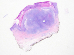
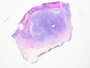
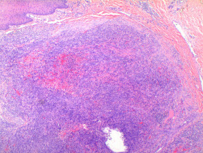
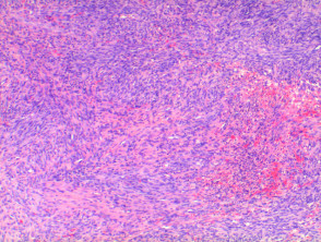
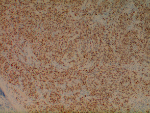
Viewed at low power, the histology of Kaposi sarcoma reveals a dermal cellular dermal nodule (Figure 1). Varying degrees of superimposed epidermal alterations can be seen, epidermal cells ranging from prominent hyperkeratosis y acanthosis hyperkeratosis and acanthosis to frank ulceration ulceration. The proliferation in the dermis consists of a growth of spindle cells; specifically, endothelial cells endothelial cells that generate tortuous vascular spaces. These elements may be scarce in the earliest lesions lesions in the patch stage, evolving into fascicles of spindle cells fascicles of spindle cells in nodular lesions (Figures 2 and 3). This fascicular pattern is often described as resembling a fish school.
The spindle cells will infiltrate the collagen, collagen, creating slit-like spaces, particularly toward the outer borders of the lesions lesions (Figure 4). When newly formed vessels penetrate a pre-existing space, the cobblestone sign sign (Figure 5) is observed, although it is now recognized that this finding is not exclusive to this pathology.
In plaque and nodular stages, it is common to find hyaline globules hyaline fat both intracellular intracellular as well as extracellular, which typically represent phagocytosed erythrocytes erythrocytes. Although infrequent, this phenomenon can be seen even in patch lesions and PAS stains are positive. It is important to note that, while the endothelial proliferation often maintains a monomorphic appearance, monomorphic, marked evidence of nuclear atypia significant (See Figure 5). RNA accompanied by an increase in mitotic activity mitotic (Figure 6). When tumor atypia is very marked, the tumor tumor may be classified as an anaplastic variant..
The inflammatory infiltrate inflammatory is predominantly lymphocytic lymphocytic, supplemented by the presence of plasma cells plasma scattered, this being a useful indicator for diagnosis. Erythrocytes are commonly observed within the slit spaces and disseminated throughout the tumor. Occasionally, at the base or periphery of the Kaposi nodule, large cavernous vessels may be identified.
Kaposi Sarcoma Pathology Gallery

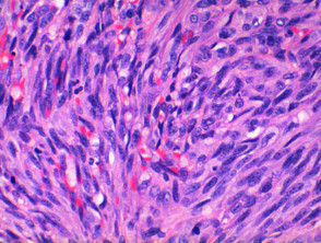

Understanding the progressive histopathological characteristics of Kaposi sarcoma, from subtle patch changes to the complex morphology of nodules, is fundamental for accurate diagnosis. The correct identification of endothelial spindle cells and the evaluation of atypia allow for the distinction of this vascular neoplasm and the determination of its corresponding clinical variant.
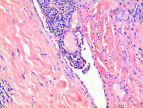
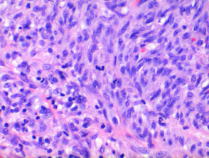
Histological Variants of Kaposi Sarcoma
Numerous pathological variations pathological have been identified for Kaposi sarcoma, which are defined according to associated additional features or the specific pattern of proliferation in the dermis. These classifications are generally deductive from the morphological description of the lesion. Reported histological variants include:
- Anaplastic
- Telangiectatic
- Lymphedematous
- Hyperkeratotic
- Keloid
- Micronodular
- Pyogenic granuloma-like granuloma differential diagnosis
- Ecchymotic
- Intravascular
Specialized Diagnostic Tests for Kaposi Sarcoma
Kaposi sarcoma lesion cells express nonspecific endothelial markers, such as CD31 (Figure 7), and CD34, in addition to the lymphatic endothelial marker system. D2-40 or podoplanin. The antibody directed against latent nuclear antigen antigen RNA latency period 1 of HHV-8 (human herpesvirus 8) constitutes a highly specific staining technique that has optimized diagnosis in complex clinical cases (Figure 8).
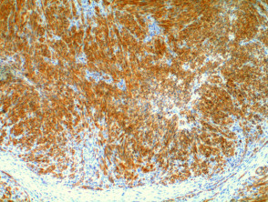

The definitive diagnosis of Kaposi sarcoma, especially in its less common variants or in early stages, is fundamentally supported by the correlation of clinical findings with histopathological analysis and the described immunohistochemical tests. The precise identification of markers such as CD31 and specific staining for HHV-8 are crucial steps to confirm the neoplastic and etiologic nature of the lesion.
Differential Diagnosis of Kaposi Sarcoma
The differential diagnosis of Kaposi sarcoma (KS) requires careful distinction, especially in its initial stages or when lesions present atypically. The following vascular conditions should be considered:
- Microvenular Hemangioma: This vascular tumor presents a diagnostic challenge against early patch lesions of KS. If less defined vessels, a significant degree of cellular pleomorphism, and positive immunohistochemistry for HHV-8 are observed, the diagnosis leans toward Kaposi sarcoma.
- Tufted Angioma: Although it shows multiple lobules composed of spindle cells, this type of hemangioma is distinguished from KS by the absence of the typical interlacing pattern and the formation of slit-like vascular channels characteristic of the latter.
- Pseudo-Kaposi Sarcoma (Acroangiodermatitis): In the differential diagnosis of early KS lesions, acroangiodermatitis is characterized by an organized proliferation of small vessels, lacking the sinuous and proliferative appearance observed in Kaposi sarcoma.
- Cavernous Hemangioma: This entity should be considered if nodular KS lesions present dilated vessels at their periphery or base. However, cavernous hemangioma does not exhibit the typical spindle cell component of KS.
- Pyogenic Granuloma: Generally, the diagnosis is complicated only when the biopsy is incomplete, showing only the superficial component of the lesion, which can simulate the vascular characteristics of KS.
Correct differentiation is crucial for establishing the appropriate therapeutic management of Kaposi sarcoma.


