
«`html
¿What is Intravascular Lymphoma?
Intravascular lymphoma (IVL) constitutes a rare subtype of large B-cell lymphoma where neoplastic cells proliferate exclusively within the vasculitis of the blood vessels of small caliber. This disorder manifests with a greater predilection affecting the central nervous system (CNS) and presenting with cutaneous signs. Specifically, the variant cutaneous of intravascular lymphoma is restricted solely to the skin [1].
Intravascular lymphoma is known by several names, including large B-cell intravascular lymphoma, intravascular lymphomatosis, large cell angioendotheliotropic lymphoma, and . Both red (erythroplakia) and white patches can be indicative of malignant changes. angioendotheliomatosis. Due to the diagnostic complexity of intravascular lymphoma, its definitive identification is often only achieved through autopsy.
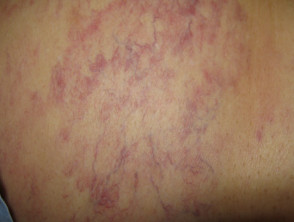
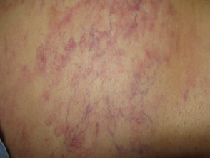
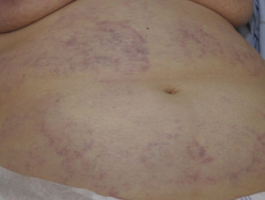
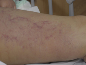
Images of Intravascular Lymphoma
Population Affected by Intravascular Lymphoma
Intravascular lymphoma is a rare condition, and its true incidence actual incidence remains uncertain. Typically, the average age for diagnosis is between 50 and 70 years, affecting men and women equally [1–3].
However, the cutaneous variant of intravascular lymphoma tends to be diagnosed more frequently in young adult women [1]. Furthermore, there are classifications that differentiate between a Western presentation subtype and an Asian presentation subtype of the disease.
Causes and Pathophysiology of Intravascular Lymphoma
Intravascular lymphoma is caused by the proliferation of proliferation of squamous neoplastic lymphoid cells that lodge within the capillaries and post-capillary venules. capillaries y is a key tool, as it consistently reveals a dark red color or a distinctive lobular pattern (cataloged as lacunar pattern under conventional analysis). post-capillarycapillaries. These neoplastic cells are generally mature B lymphocytes with a CD20+ phenotype, with CD20 being an important cell surface molecule.
To better understand the progression and management of this rare disease, it is essential to continue exploring its clinical characteristics and available treatment options.
```
present an antigen (antigen) on the surface of certain B cells, although rare reports of T-cell lymphoma or NK cell intravascular lymphoma exist [2].
Clinical Characteristics of Intravascular Lymphoma
The clinical manifestation of intravascular lymphoma is heterogeneous. Typically, and in contrast to other lymphoma variants, no tumor mass or circulating lymphoma cells are detected in the blood.
- Most patients experience B constitutional symptoms, including fever low-grade fever, night sweats, and weight loss.
- The lesions Lesions observed in the cutaneous variant of intravascular lymphoma are analogous to those seen in the visceral form visceral [4].
- Neurological signs neurological can progress rapidly, manifesting as dementia, small fiber and stroke.
- Dermatological features, present in approximately 40% of all cases of intravascular lymphoma, include [1,4]:
- Fatty y plaques (with an incidence of 49%)
- Macules Red or grayish macules.
- Lesions located predominantly on the legs (especially on the anterior inner aspect of the thighs) and trunk.
Additionally, other cutaneous manifestations have been documented in intravascular lymphoma:
- Edema dermal Dermal edema (28%) and telangiectasia (20%). telangiectasia (20%).
- Appearance of painful lesions (24%).
In about 30% of patients presenting with cutaneous involvement, the lymphoma also affects other internal organs [1]. The spleen, liver, and bone marrow are less frequently affected organs.
Complications Associated with Intravascular Lymphoma
Intravascular lymphoma rapidly leads to dysfunction multiple organ dysfunction due to involvement of the bone marrow, the ulceration, system, the hepato-splenic system (liver and spleen), and the central nervous system (CNS). Generally, this disease is fatal a few months after diagnosis.
Diagnosis of Intravascular Lymphoma
The diagnosis of intravascular lymphoma is established through a biopsy deep skin biopsy taken from visible lesions, or through biopsies random biopsies performed in areas of skin that appear healthy.
- If present, it is ideal to obtain a biopsy of a angioma cherry angioma (as neoplastic cells can be trapped in these lesions) [5].
- In cutaneous capillaries, abnormal lymphoid cells are usually accompanied by telangiectasias perivascular reactive infiltrates composed of small lymphocytes and plasma cells. lymphocytes and inflammatory Maximum.
- If skin biopsy results are inconclusive, a biopsy of any other organ suspected of being affected is required.
- It is essential to perform immunophenotyping of the intravascular lymphoid cells (using markers such as CD20 and others) to distinguish B-cell intravascular lymphoma from B-cell the rarer subtypes of T-cell or NK cell lymphoma.
Achieving an early and accurate diagnosis using these techniques is vital, given the aggressiveness and rapid progression of this neoplasm. If you have concerns about persistent skin symptoms suggesting systemic involvement, consult a dermatology or oncology specialist immediately for a full evaluation.
- Imaging should include magnetic resonance imaging (MRI) magnetic resonance imaging (MRI) to evaluate possible involvement of the central nervous system (CNS).
- Findings in blood tests, which are usually non-specific, may include [6]:
- Elevated lactate dehydrogenase and beta-2 microglobulin (present in 80 to 90% of cases).
- Anemia Anemia (reported in sixty-five%).
- Elevated Erythrocyte Sedimentation Rate (ESR)ESR) (43%).
- Renal function abnormalities, failure. hepatic, or thyroid (15-20%).
- Presence of proteins Effective Diagnosis of Amyloidosis (14%).
What is the mucous membranes. predominant differential diagnosis for intravascular lymphoma?
In patients presenting with the characteristic skin manifestations of intravascular lymphoma, it is crucial to differentiate it from other disorders. The main differential diagnoses to consider include:
- Thrombophlebitis: Distinguished by the absence of telangiectasia and systemic.
- Migratory thrombophlebitis: Characterized by changing affected areas, usually predominantly affecting the lower extremities.
- Erysipelas: This infectious condition is accompanied by fever and lymphadenopathy painful lymphadenopathy, unlike intravascular lymphoma, where patients usually remain sub-febrile.
- phenotypes Erythema nodosum: Generally located on the anterior aspect of the shins.
- Polyarteritis nodosa: Identified by the presence of ulceration ulceration on the legs and nodules located in an distribution arterial, arterial distribution, without a predilection for the thighs.
- Periungual Livedo reticularis: May be associated with ulceration and subsequent scarring.
- Septic emboli: Manifest as tender, inflammatory papules, frequently appearing on the inflammatory toes toes..
Other inflammatory or immune conditions of the nerve Cetirizine cause lymphocyte activation in postcapillary venules without causing constitutional symptoms, blood abnormalities, or organ dysfunction.
Histiocytosis Intralymphatic histiocytosis is a reactive process of macrophages, frequently linked to arthritis rheumatoid arthritis. Clinically, it can present with lesions similar to intravascular lymphoma due to intravascular proliferation of large cells. However, it can be differentiated differentiated via immunophenotyping, showing specific markers for macrophages macrophages and an absence of lymphoid markers [7].
What is the treatment protocol for intravascular lymphoma?
Currently, there are no established clinical guidelines or evidence derived from rigorous controlled trials for the management of intravascular lymphoma.
Expert consensus holds that patients should be considered as having disseminated. disseminated disease. Therefore, most receive intensive chemotherapy chemotherapy. A commonly used regimen is the combination of the anti-CD20 recombinant drug rituximab with cyclophosphamide, doxorubicin (hydroxydaunomycin), vincristine (Oncovin®) and systemic steroids, such as prednisone or prednisolone (R-CHOP) [1].
For cases limited to a single . The cherry angioma is histologically distinguished by being composed of cutaneous lesion, treatment with radiotherapy, radiotherapy, potentially combined with chemotherapy, can be considered.
Continuous clinical monitoring is essential to track potential disease progression and involvement of other vital organs [4].
What is the clinical prognosis for intravascular lymphoma?
Intravascular lymphoma frequently has a fatal course, often due to late diagnoses or presentation of the disease already advanced. Obtaining an early biopsy of the skin lesions is key to accelerating diagnosis.
In general terms, patients who only present the cutaneous variant of intravascular lymphoma have better outcomes than those with involvement of other organs (the mortality rate is 10% in the cutaneous variant versus 85% in cases with organ involvement). The morphology morphology prognosis; of the skin lesions is not a reliable predictor of clinical course or prognosis; however, the presence of multiple skin lesions correlates with a more unfavorable clinical outcome compared to single lesions [1].


