
Understanding Granulomatous Dermatitis: Types and Key Characteristics
Granulomatous dermatitis encompasses various skin conditions distinguished by their histological particularity histological.
- Interstitial Granulomatous Dermatitis (IGD)
- Palisaded Neutrophilic Granulomatous Dermatitis (PNGD)
- Granulomatous Interstitial Reaction to Drugs (IGDR)
Interstitial Granulomatous Dermatitis (IGD)
Interstitial Granulomatous Dermatitis is a rare dermal disorder, characterized by a specific pattern of inflammation in the skin. inflammation Macular amyloidosis.
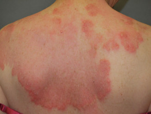
The original and most classic clinical description of IGD involved the appearance of palpable, linear erythematous cords cords with elevated palpable y linear on the flanks of the trunk, which was termed the «string sign» (sign). However, numerous types of rashes sharing the same histological presentation have since been identified.
Clinical Characteristics of Interstitial Granulomatous Dermatitis
The clinical manifestations of Interstitial Granulomatous Dermatitis exhibit notable variability:
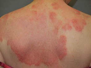
- Presence of reddish or skin-colored macules or patches, along with papules y plaques.
- The morphology of the lesions The morphology of the lesions, annular can be round, annular (ring-shaped), or have a cord-like appearance. cord-like.
- The lesions are fluctuating, increasing and decreasing in size and shape over days or months.
- They are generally asymptomatic, although some patients report mild pruritus (itching) or a burning sensation.
- They tend to be distributed symmetrically on the trunk, but the proximal extremities proximal can also be affected.
- It occurs most frequently in middle-aged women.
- A significant proportion of diagnosed patients also present with some concomitant autoimmune disease. defect concomitant.
Interstitial Granulomatous Dermatitis
Interstitial Granulomatous Dermatitis
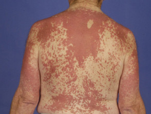
Interstitial Granulomatous Dermatitis
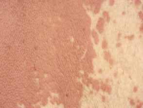
Interstitial Granulomatous Dermatitis
Diagnosis of Interstitial Granulomatous Dermatitis
The definitive diagnosis of Interstitial Granulomatous Dermatitis is established through histopathological analysis of a skin biopsy biopsy performed by a pathologist pathologist. The distinctive histological features that confirm IGD include:
- A dense histiocytic inflammation lymphadenitis located in the reticular dermis reticular (lower layer).
- The presence of an infiltrate with sparse neutrophils.
Understanding these specific histological criteria is fundamental, as the clinical appearance can overlap with other conditions. If you suspect a skin disorder, it is crucial to consult a specialist for an accurate diagnosis and to determine the most appropriate therapeutic approach to manage the granulomatous dermatitis.
- Notable presence of eosinophils.
- Infiltrate perivascular and interstitial lymphocytes.
- Histiocytes interstitial (located between the collagen bundles) collagen) or palisaded histiocytes (aligned perpendicularly to fragmented central collagen).
- Focal degeneration of collagen, which may be surrounded by a space (floating sign).
- Few giant cells.
Differences Between Interstitial Granulomatous Dermatitis and Granuloma Annulare
Granuloma annulare and interstitial granulomatous dermatitis can present with similar clinical and histological pictures. histological similar. Granuloma annulare typically manifests with papules and plaques localized on the dorsa of the hands or feet, whereas interstitial granulomatous dermatitis more frequently affects the trunk as the primary area.
Furthermore, granuloma annulare is less frequently associated with autoimmune pathologies.
The histological findings that characterize granuloma annulare include:
- Histiocytes positioned in the upper dermis.
- Rare or no presence of neutrophils and eosinophils.
- Abundance of mucin [1].
Palisaded Neutrophilic Granulomatous Dermatitis: Characteristics and Diagnosis
Palisaded neutrophilic granulomatous dermatitis was initially identified by its crusted and umbilicated papules umbilicated on the elbows, presenting in patients with arthritis rheumatoid arthritis (known as Winkelmann's Granuloma), or in cases of eosinophilic granulomatosis with polyangiitis (Churg-Strauss Syndrome, eosinophilic syndrome)., syndrome eosinophilic).
Although the histological appearance is consistent, various types of associated exanthems have been documented, including annular plaques localized on the trunk. These lesions are usually tender to the touch and have the potential for ulceration.
Histopathological Diagnosis of Palisaded Neutrophilic Granulomatous Dermatitis
The definitive diagnosis of palisaded neutrophilic granulomatous dermatitis is established via a skin biopsy. The characteristic histological hallmarks of this condition include:
- A marked neutrophilic infiltrate and presence of nuclear dust. RNA.
- Interstitial histiocytic infiltrate.
- Evidence of collagen degeneration.
- Vasculitis leukocytoclastic vasculitis..
- The histological pattern may vary depending on the evolutionary stage of the eruption. rash.
toxicity granulomas Palisaded granulomas have also been described in the literature as «miniature Churg-Strauss granulomas» or flame figures, where degenerated collagen is surrounded by eosinophils, resembling a flame. Similar phenomena are observed in the pathology pathology of Wells Syndrome.
Common Clinical Associations with Granulomatous Dermatitis
Various conditions have been documented in conjunction with interstitial granulomatous dermatitis. However, information regarding specific clinical associations for palisaded neutrophilic granulomatous dermatitis is more limited.
Associated Conditions and Diseases
Autoimmune Associations of Granulomatous Dermatitis
Both presentations of granulomatous dermatitis frequently emerge in individuals with other conditions considered autoimmune, suggesting that an immune complex mechanism might be involved in the [1,3]. [2]. These associations include the following conditions:
- Rheumatoid and non-rheumatoid arthritis, characteristically symmetrical and involving the fingers, wrists, elbows, and shoulders, constitutes the most common comorbidity. Arthritis (joint inflammation) or arthralgia (joint pain) may precede, accompany, or appear years after the onset of skin lesions in about 50% of published cases of interstitial granulomatous dermatitis [3].
- Systemic lupus erythematosus systemic (SLE).
- Antiphospholipid Syndrome primary.
- Systemic lupus erythematosus (SLE).
- Thyroiditis.
- Vitiligo.
In a literature review covering 15 patients with interstitial granulomatous dermatitis, the autoantibodies autoantibodies identified in blood tests included rheumatoid factor (RF), antinuclear factor (ANA), thyroglobulin, histone SS-A, DNA histone, and ANCA [4].
Association with Malignancy
There are several reports associating interstitial granulomatous dermatitis with malignant processes. In one documented case, the skin lesions disappeared after treatment for cancer lung cancer [5]. Uncommon associations of granulomatous dermatitis with leukemia [6], lymphoma, leukemia [6], lymphoma, breast cancer, carcinoma of squamous cells hypopharyngeal squamous cell carcinoma, and neoplasia endometrial neoplasia have been reported.
Treatment of Granulomatous Dermatitis
Granulomatous dermatitis frequently runs a course of flares and remissions. Among the treatments that have proven effective are:
- Systemic topical.
- Hydroxychloroquine.
- The selection of the most appropriate treatment should be determined by a healthcare professional, considering the intensity of the symptoms and the patient's general health.
A case associated with SLE responded successfully to systemic steroids after 15 days [2].
Although and Tumor necrosis factor inhibitors necrosis tumoral (TNFtumor necrosis factor (TNF)-alpha inhibitors have recently been described as inducers of interstitial granulomatous dermatitis [7], etanercept has been used in interstitial granulomatous dermatitis linked to rheumatoid arthritis, achieving complete skin clearance and improvement of associated arthritis [8].
Granulomatous Interstitial Reaction to Drugs (Drug Reaction with Interstitial Granulomatous Reaction)
When interstitial granulomatous dermatitis is triggered by medications, it is called a granulomatous interstitial reaction to drugs. This entity is considered a distinct clinical and of melanoma and is fundamental for determining subsequent treatment steps. pathological unit. It manifests through annular plaques and nodules nodules located on the trunk, arms, the middle It is crucial to keep in mind that a notable proportion of patients (between 10% and 15%) who initially present with SCLE may progress to full Systemic Lupus Erythematosus (SLE). This progression carries the potential risk of developing serious involvement, including part of the thighs, and skin folds. The rash completely resolves after discontinuation of the causative pharmacological agent.
Diagnosis of Granulomatous Interstitial Reaction to Drugs
The diagnosis of granulomatous interstitial reaction to drugs is confirmed by a skin biopsy. The characteristic histological findings for this reaction include:
- Basal Other conditions that cause significant edema in the superficial dermis include mild cellular vacuolization.
- Basal epidermal vacuolar changes. lichenoid Lichenoid-type alterations accompanied by eosinophils.
- Absence of neutrophils in the infiltrate.
- In some cases, presence of palisaded granulomatous changes associated with collagen necrosis and the floating sign.
Among the medications reported as implicated in drug-induced granulomatous interstitial rash are:
- toxicity and TNF-alpha inhibitors (infliximab, adalimumab, etanercept) [7]
- Beta-blockers.
- Calcium channel blockers.
- Enzyme Angiotensin-converting enzyme inhibitors.
- Lipid-lowering lipids.
- agents. Statins (HMG-CoA reductase inhibitors).
- Furosemide.
- Antidepressants.
- Anticonvulsants [4].
Accurate identification of the causal agent is crucial to achieve remission of the granulomatous dermatitis and prevent recurrences. If you suspect an adverse drug reaction, consult your dermatologist or specialist immediately for a complete diagnostic evaluation.


