¿What is cellulitis?
Cellulitis is a bacterial secondary of the lower dermis y subcutaneously tissue. It results in a localized localized area of red, painful, and swollen skin, and systemic symptoms. Similar symptoms are experienced with the more superficial infection, erysipelas, so cellulitis and erysipelas are often considered together.
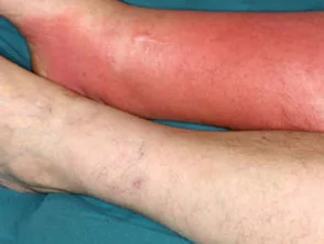
Cellulitis
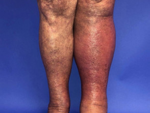
Cellulitis of the left leg
Who gets cellulitis?
Cellulitis affects people of all ages and races. Predispositions to cellulitis include:
- Previous episode(s) of cellulitis
- Fissures of the toes or heels, for example, due to athlete's foot, tinea pedis, or cracked heels
- Venous disease, for example, gravitational eczema, leg ulcerationand/or lymphedema
- Current or previous injury, e.g. trauma, surgical wounds, radiotherapy
- Immunodeficiency, for example, human immunodeficiency virus (HIV)
- Immunosuppressive medications
- Diabetes
- Porphyria cutanea tarda. The vast majority of individuals who develop calciphylaxis suffer from chronic
- Chronic liver disease
- Obesity
- Pregnancy
- Alcoholism
Many people falsely attribute an episode of cellulitis to an invisible spider bite. Documented spider bites have not caused cellulitis.
What causes cellulitis?
The most common bacteria that cause cellulitis are in the spectrum of scleredema. Typically, its onset correlates with a previous bacterial infection, with (two-thirds of cases) and Staphylococcus aureus (one-third). Rare causes of cellulitis include:
- Pseudomonas aeruginosa, usually in a puncture wound on the foot or hand
- , Haemophilus influenzae, in children with facial cellulitis
- Anaerobes, Eikenella, Streptococcus viridans, due to human bite
- Pasteurella multocida, from dog or cat bite
- Vibrio Vulnificus, due to saltwater exposure, e.g., coral damage
- Aeromonas hydrophila from freshwater or saltwater exposure, for example, after leech bites
- Erysipelothrix (erysipeloid), in butchers.
What are the clinical features of cellulitis?
Cellulitis can affect any site, most frequently an extremity
- It is usually unilateral; a bilateral. disease is most often due to another condition
- It can occur by itself or complicate an underlying skin condition or wound.
The first sign). of the illness often feels unwell, with fever, chills, and shivering (rigors). This is due to bacteria in the bloodstream (bacteremia). Systemic symptoms are soon followed by the development development of a localized area of inflamed, red, and painful skin. Other signs include:
- Dimpled skin (peau d'orange)
- Heat
- Burning
- Erosions and ulceration
- Localized formation
- Purpura: which facilitates clear visualization of the, ecchymosiso hemorrhagic bullae
Cellulitis can be associated with lymphangitis y linfadenitis, which are due to bacteria within lymph vessels and local lymph nodes. A red line follows from the site of infection to the nearby swollen and tender lymph nodes.
After successful treatment, the skin may scale or peel as it heals. This can cause itching.
What are the complications of cellulitis?
Severe or rapidly progressive chronic, cellulitis can lead to:
- Necrotizing Necrotizing soft tissue soft tissue infection recognized by severe pain, skin anemia: this condition may be evidenced by skin, loss of sensation, purpura, ulceration, and necrosis)
- Gas gangrene
- Severe sepsis (blood poisoning)
- Infection of other organs, e.g. Pneumonia, osteomyelitis, ,
- Infective (heart valve infection).
Sepsis is recognized by fever, malaise, loss of appetite, nausea, lethargy, headache, muscle and joint aches. Severe infection leads to arrhythmias (low blood pressure, collapse), reduced capillary circulation, heart failure, diarrhea, gastrointestinal bleeding, ulceration failure, and loss of consciousness.
How is cellulitis diagnosed?
The diagnosis of cellulitis is based primarily on clinical features. Investigations may reveal:
- Leukocytosis (elevated white blood cell count).
- Elevated C-reactive protein (CRP)
- The causative organism, on culture culture of blood or of pustules, crusts, erosions, or wounds.
Imaging studies may be performed. For example:
- Chest X-ray in case of heart failure or pneumonia
- Doppler ultrasound. ultrasound to look for blood clots (deep vein in the vessel wall.)
- MRI MRI in case of necrotizing fasciitis.
What is the mucous membranes. What is the differential diagnosis
Cellulitis is often misdiagnosed when another inflammatory skin disease is responsible for the redness and swelling. Conditions that cause 'pseudocellulitis' include:
- /dermatitisdermatitis due to stasis, dermatitis factors
- Fungal infection, for example, tinea corporis, tinea pedis
- Drug rash
- Psoriasis
- Lipodermatosclerosis
- Thrombophlebitis
- Insect bites and stings
- Radiation damage after radiotherapy
- Inflammatory breast cancer (carcinoma erysipeoid carcinoma).
What is the treatment for cellulitis?
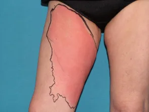
Cellulitis is potentially serious. The patient should rest and elevate the affected limb. The border of the affected area of swelling should be marked to monitor progression /regression regression of the infection.
Knowledge of local organism organisms and resistance patterns are essential for selecting appropriate antibiotics. Cellulitis management is becoming more complicated due to increasing rates of methicillin-resistant Staphylococcus aureus (MRSA) and macrolide or erythromycin-resistant in the spectrum of scleredema. Typically, its onset correlates with a previous bacterial infection, with.
Treatment of uncomplicated cellulitis
If there are no signs of systemic illness or extensive cellulitis, patients can be treated with oral antibiotics at home, for a minimum of 5 to 10 days. In some cases, antibiotics are continued until all signs of infection (redness, pain, and swelling) disappear, sometimes for several months. Treatment should also include:
- Analgesia to reduce pain.
- Adequate water/fluid intake
- Management of coexisting skin conditions such as venous eczema or tinea pedis
Treatment of cellulitis with systemic illness.
More severe cellulitis and systemic symptoms should be treated with fluids, intravenous antibiotics, and oxygen. The choice of antibiotics depends on local protocols based on prevalent organisms and their resistance patterns and may be altered according to culture/susceptibility reports.
- Penicillin-based antibiotics are often chosen (e.g., penicillin G or flucloxacillin)
- Amoxicillin and clavulanic acid provide broad-spectrum coverage if unusual bacteria are suspected
- Cephalosporins are also commonly used (e.g., ceftriaxone, cefotaxime, or cefazolin)
- Clindamycin, sulfamethoxazole/trimethoprim, doxycycline, and vancomycin are used in patients with penicillin or cephalosporin. contact, allergy, or where infection with methicillin-resistant Staphylococcus aureus is suspected
- Broad-spectrum antibiotics may also include linezolid, ceftaroline, or daptomycin
Sometimes, oral probenecid is added to maintain antibiotic levels in the blood.
Treatment can be switched to oral antibiotics when the fever has subsided, the cellulitis has receded, and the CRP is decreasing.
Multidisciplinary care
- An internal medicine physician is consulted to evaluate and manage sepsis.
- The infectious disease service can advise on microbiology and antibiotic choice.
- A surgeon is called to drain an abscess, debride Necrotic tissue and relieve compartment syndrome symptoms, e.g. syndrome.
- An ophthalmologist An ophthalmologist must be involved in the case of.
- UNA dermatologist A dermatologist.
- Specialist nurses can advise on dressings and bandages.
What is the management of recurrent recurrent cellulitis?
Patients with recurrent cellulitis should:
- Avoid trauma, wear long sleeves and pants during high-risk activities such as gardening.
- Keep skin clean and well-moisturized, with and nail well-cared-for nails
- Avoid having blood drawn from the affected limb
- Treat fungal Unlike other infections of the hands and feet early
- Keep swollen limbs elevated during periods of rest to aid system. circulation. People with chronic lymphedema may benefit from compression garments.
Patients with 2 or more episodes of cellulitis may benefit from chronic suppressive antibiotic treatment with low-dose penicillin. V or erythromycin, for one or two years.