
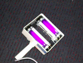
Wood's Lamp Examination for Skin Diagnosis
The Wood's lamp examination is a specialized diagnostic technique where the skin or hair hair is evaluated under exposure to the black light emitted by this specific lamp. The black light is invisible to the human eye as it operates in the ultraviolet spectrum, with a wavelength just shorter than the color violet. The lamp usually emits a violet glow in dark environments, as it also projects a small portion of light in the violet section of the electromagnetic spectrum.
What Instrument is the Wood's Lamp?
The traditional tool, the Wood's lamp, consists of a low-power mercury arc covered by a Wood's filter (composed of barium silicate and 9% nickel oxide), which allows it to emit a wavelength between 320 and 450 nm (with a peak around 365 nm). This device was conceived in 1903 by the physicist Robert W. Wood, originally from Baltimore.
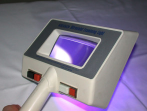
Today, modern black light sources include specially designed BLB fluorescent lamps, mercury vapor lamps, light-emitting diodes (LEDs), or even incandescent lamps. Fluorescent black light tubes use a dark blue filter coating that suppresses most of the visible light. Wood's lamps used in the medical field may incorporate:
- Various phosphors that present different emission peaks.
- Magnifying lenses for better visualization.
- A white light option.
- A black curtain to completely eliminate ambient light.
Procedure for the Wood's Lamp Skin Examination
The evaluation process using the Wood's lamp follows these key steps.
- Ideally, the skin area to be examined should not have been recently washed or have cosmetics, deodorants, or lotion, as these products can induce fluorescence and generate a false-positive result.
- Gentle facial cleansing may be required beforehand.
- The Wood's lamp is connected and allowed to warm up for approximately one minute to reach its optimal performance.
- Ambient lights are turned off and window curtains are closed, or dark curtains are used to achieve total dimness in the environment.
- After allowing the eyes to adapt to the dark, the skin is observed with the Wood's lamp for a few seconds. The instrument is kept at a safe distance, between 10 and 30 cm from the skin surface. This procedure is painless and considered completely safe.
This lamp is used to determine the extent of areas with pigmented alterations or pigmented or depigmented areas, and to detect abnormal fluorescence patterns. Healthy, normal skin may show a slight bluish tint, but will present white areas in thickened zones, yellowish if oily, and purple where there is dehydration. Additionally, clothing fibers often glow an intense white under this light.
A result is considered positive if a pigmentary disorder pigmentation becomes significantly more visible when examined with the Wood's lamp, or if any striking fluorescence is observed.
Wood's lamp
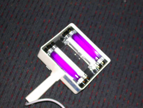
Burton Wood Lamp
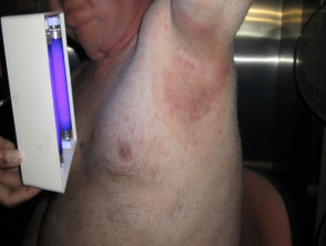
What is Fluorescence in Dermatology?
Fluorescence is defined as the optical phenomenon in which certain substances emit a visible color glow. This occurs when specific materials, such as collagen and porphyrins, collagen and Urinary, fecal, or serum, absorb ultraviolet light (or black light) and immediately re-emit it at a longer wavelength that falls within the visible spectrum. Additionally, compounds present on the skin surface, such as soap residue, certain topical medications, or textile fibers, can also show this fluorescent effect under Wood's light.
Dermatological Conditions Diagnosed with Wood's Lamp
The Wood's lamp examination is a fundamental diagnostic tool that helps reveal and differentiate various skin conditions by observing specific fluorescence patterns. The lamp can identify:
- Increased Pigmentation: It is used to determine if hyperpigmentation (such as melasma or post-inflammatory pigmentation) is of epidermal (where pigmentation intensifies notably under Wood's light) or dermal (where pigmentation shows no clear changes). Pigmented lesions exhibit a defined border under ultraviolet light due to light absorption by excess melanin. melanin.
- Loss of Pigmentation (Hypopigmentation): It is useful for outlining affected areas in cases such as vitiligo, macules Lesions of the systemic sclerosis tuberous sclerosis, and hypomelanosis of Ito, especially in fair-skinned patients. Hypopigmented skin exhibits sharper borders and an intense blue-white fluorescence (or sometimes yellowish-green) resulting from the accumulation of biopterins. In contrast, areas with reduced blood flow show no change.
- Tinea Versicolor: This rash persistent granular scaly, frequently located on the chest and upper back, is caused by the yeast *Malassezia*. When active, the scale The presence of a superficial produces a characteristic yellow or orange glow.
- Malassezia Folliculitis: toxicity follicles Hair follicles infected by this yeast present a blue-white fluorescence.
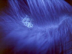
- Tinea Capitis: This secondary fungal infection causes areas of scaling and alopecia. Species of the genus *Microsporum* (*M. canis*, *M. audouinii*, *M. ferrugineum*, *M. distorsum*) emit a blue-green fluorescence, while *Trichophyton schoenleinii* produces a dull blue tone. Infections caused by other organism do not usually fluoresce.
- Infestations: Detects the presence of lice and scabies burrows.
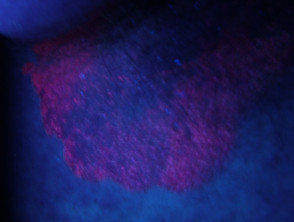
- Erythrasma: The bacteria The genus *Corinebacterium*, responsible for this pigmented rash in skin folds, emits a coral pink color under Wood's light.
- Pseudomonas Infections: The presence of *Pseudomonas* in jacuzzi-associated folliculitis or in infected wounds produces a green light.
- Acne Vulgaris: The accumulation of *Cutibacterium* (formerly *Propionibacterium*) in hair follicles causes acne to turn orange-red.
- Porphyrias: It causes a reddish-pink fluorescence on the skin (in the case of porphyria cutanea tarda) or on the teeth (in erythropoietic porphyria).
- Photodynamic Therapy: The porphyrins used as photosensitizers in photodynamic therapy photodynamic are visible.
- Drug Reactions: Certain ingested medications, such as tetracyclines and mepacrine, may fluoresce after ingestion.
- Chemical Peel Monitoring: It evaluates the uniformity in the application of solutions containing salicylic acid, which turns fluorescent green upon contact.
Observation of Fluorescence During the Wood's Lamp Examination
Diverse Applications of the Wood's Lamp
In addition to skin diagnosis, the Wood's Lamp offers valuable utilities in multiple specialized fields and laboratories.
- In the biochemistry laboratory, it is used to identify traces of porphyria, detecting erythropoietic protoporphyrin in red blood cells, or observing porphyria cutanea tarda in urine and stool samples.
- The molecular biology laboratory can use the Wood's lamp to visualize compounds labeled with fluorescent tags.
- Estheticians use it to evaluate early signs of skin aging and locate other dermal imperfections.
- Ophthalmology professionals use it to discover scratches or identify foreign bodies lodged in the infiltrates of the eye.
- Veterinary surgeons use this tool when examining pets for bacterial infections, bacterial, fungal, or parasitic infections.
- Law enforcement uses it to examine crime scenes and articles of clothing in the forensic search for traces of semen or urine.
- It is effective for locating pet urine or feces stains Perform checks and adequately clean clogged gutters and flat roof surfaces where water can stagnate. on surfaces.
- Industrial evaluation of metal fatigue can be performed by applying a fluorescent fluid and subsequently examining it with the Wood's lamp.
- In recreational settings, nightclubs use it to verify the authenticity of fluorescent re-entry stamps.
- Immigration officers use it to certify the authenticity of passports.
- Banking institutions use it to detect counterfeit bills.
- In geology, it is fundamental for mineralogy and gemology.
- It is used to display artwork created with fluorescent paint in art and sculpture.
Does the Wood's Lamp Pose Any Health Risk?
The black light emitted by the Wood's Lamp is intrinsically harmless. This device does not produce short-wave ultraviolet B radiation (range of 290–320 nm); therefore, it has no potential to cause sunburn or damage healthy skin tissue.
In rare cases, a patient with a condition of extreme reactions. may experience the appearance of a rash in the area exposed to black light. However, since the Wood's Lamp examination is generally very quick, complications are highly unlikely, even in dermatoses..
As a standard and prudent precautionary measure, it is recommended to ask the patient to keep their eyes closed while the facial inspection is performed.


