
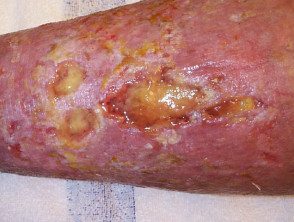
Definition and Causes of Venous Stasis Ulcer
A stasis ulcer represents the most frequent form of chronic full-thickness skin loss localized to the lower leg. This condition gets its name because it involves an incompetence in the venous system, which prevents the adequate flow of venous blood from the lower extremities back to the heart. It is important to note that the stasis ulcer is also known as a lower leg venous ulcer or gravitational ulcer; however, the term varicose ulcer is inappropriate if associated varicose veins are not present.
Who is susceptible to developing stasis ulcers?
Stasis ulcers are a relatively common condition, affecting approximately 1% of the population. Their development is linked to a combination of underlying medical factors and lifestyle habits. Among the main contributors are:
- Advancing age.
- History of deep vein thrombosis (DVT), which is the formation of a clot within the deep veins.
- Obesity condition.
- Previous injuries, surgeries, or fractures in the lower extremities.
- Prolonged periods of immobility.
- Hypertension (high blood pressure).
- Standing for extended periods of time.
- Having had multiple pregnancies.
- Presence of varicose veins.
These ulcers constitute at least 40-50% of all chronic lower extremity ulcers, and they also contribute to about 20% of cases involving combined arterial and venous components.
What is the etiology of stasis ulcers?
The primary origin of stasis ulcers is attributed to a combination of hemodynamic factors:
- Venous insufficiency, characterized by inefficient return of peripheral venous blood to the central circulatory system.
- Venous hypertension, which implies sustained elevated pressures within the venous system.
- Edema, which is the excessive accumulation of fluid in the tissues of the lower extremities.
Various problems directly contribute to this venous insufficiency:
- Obstruction of venous flow, caused either by DVT or obesity.
- Incompetence or damage to the valves present within the blood vessels.
- Deterioration of the "venous pump," the mechanism that relies on muscle contractions to propel venous return.
When efficient blood return to the heart is compromised, a cascade of pathological events is triggered:
- Interstitial fluid filters out from the overloaded blood vessels into the surrounding tissues.
- Vessel walls become fibrotic, meaning thickened.
- A reduction occurs in the supply of essential oxygen and nutrients to the affected tissues.
- The capacity to eliminate metabolic waste products decreases.
- Tissues experience chronic inflammation.
- The natural wound healing process becomes hindered.
The final result of this congestion and tissue damage is the erosion and subsequent breakdown of the skin, a process often precipitated after an acute injury, which may even be quite minor in nature.
Characteristic Clinical Manifestations of Stasis Ulcers
Venous stasis ulcers exhibit a distinctive set of clinical features that facilitate their identification:
-
Preferred location: They usually appear on the lower legs, most frequently in the inner ankle region or in the typical 'stocking' area.
-
Pain level: They generally cause minimal or mild pain.
-
Appearance: They tend to be superficial and are characterized by the presence of a significant amount of exudate (liquid discharge).
Exploring More About Lower Leg Stasis Ulcers
Understanding these parameters is crucial for differential diagnosis and the effective implementation of treatments for venous stasis ulcers.
Lower Leg Venous Ulcer
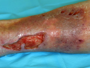
Lower Leg Venous Ulcer
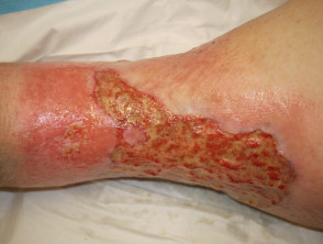
Lower Leg Venous Ulcer
Clinical manifestations that help confirm the diagnosis of venous disease include a series of cutaneous and structural signs in the lower extremities:
- Significant ankle edema.
- Presence of dilated varicose veins.
- Changes in skin pigmentation, specifically a reddish-brown coloration caused by hemosiderin deposition. released after the breakdown of red blood cells.
- Dermatitis, which can manifest as a weeping and blistering condition, or as dry, scaly, and itchy skin.
- Development of lipodermatosclerosis, characterized by thickening and hardening of the underlying tissues.
- Thickened nails that exhibit slow growth.
Complications Associated with Venous Insufficiency
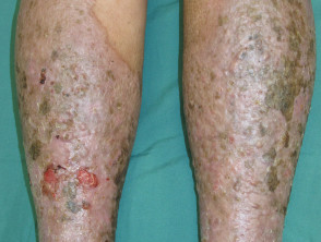
Dermatitis and Venous Ulcerations
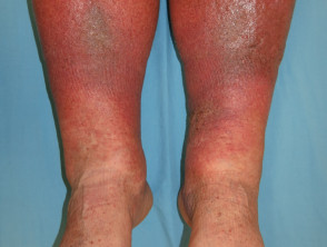
Lipodermatosclerosis
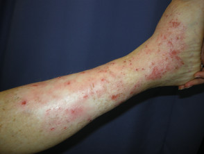
Venous Eczema
Primary Risks and Complications of Stasis Ulcers**
The most significant complication associated with stasis ulcers is the development of an secondary. This infection can seriously compromise the natural wound healing process. Although bacteria bacteria colonize virtually all leg ulcers, systemic antibiotics are not necessary unless there are clear clinical manifestations of active infection. Key indicators that a stasis ulcer might require antibiotic intervention include:
- Signs of erythema (redness) and noticeable swelling in the skin surrounding the lesion.
- Increased sensation of warmth in the affected area.
- Intensification of localized pain.
- Progressive increase in ulcer size.
- Increase in secretion. Increase in purulent discharge or tissue or oozing from the wound.
- Presence of fever Arterial insufficiency.
It is essential to note that routine antibiotics are currently not recommended, as they can slow the overall rate of wound healing, even despite their bactericidal properties. bactericidal.
Cellulitis and Its Relationship with Venous Insufficiency
When venous ulcers develop cellulitis (a serious bacterial infection of the skin and subcutaneous tissue), management becomes critical. This condition exacerbates inflammation and the risk of progression of the underlying venous disease, making it essential to promptly identify signs of infection to initiate appropriate treatment and prevent major complications such as sepsis or permanent alteration of lymphatic and venous function.
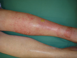
Infected Venous Ulcer
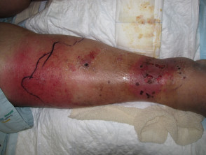
Cellulitis Around the Venous Ulcer
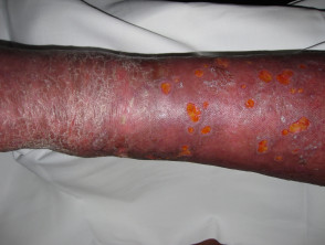
Cellulitis and Venous Insufficiency
Diagnosis and Effective Treatment of Stasis Ulcers
Diagnostic Procedures for Venous Stasis Ulcers
The diagnosis of stasis ulceration is established predominantly clinically. It is based on a detailed evaluation of the patient's medical history and findings obtained during the physical examination. This includes identifying risk factors associated with venous stasis—congestion and decreased venous circulatory flow—as well as the specific characteristics of the skin wound and surrounding tissue.
- The Duplex ultrasound is employed as a fundamental tool to confirm the existence of venous obstruction or valvular incompetence, particularly before considering a saphenous vein ablation surgery. ablation of the saphenous vein.
- The ankle-brachial index (ABI) is used to detect the presence of concomitant arterial disease, a factor that must be managed simultaneously.
To gain a deeper understanding of venous insufficiency and gather crucial hemodynamic data, complementary studies can be performed, such as:
- Venous Echo-Doppler.
- Venography or phlebography studies (ascending or descending, using contrast medium or magnetic resonance imaging techniques).
- Air venous plethysmography.
- Photoplethysmography.
- Lymphoscintigraphy.
- Liquid crystal thermography.
- Ambulatory venous pressure measurements.
- Arm-to-foot pressure difference tests.
- General venous function assessment.
- of infectious origin. disorders hematological studies clotting.
Options for Treating Stasis Ulcers
The therapeutic approach for stasis ulcers seeks to resolve the underlying etiology, provide immediate symptomatic relief, and fundamentally maximize the lesion's healing potential.
Initial Conservative Measures
- Implement weight reduction strategies in overweight or obese patients.
- Strongly recommend the patient increase physical activity to encourage better peripheral circulation.
- Control lower extremity edema (swelling) by instructing the patient to elevate the leg above heart level as frequently as possible, suggesting periods of at least 30 minutes, three times a day.
The Fundamental Importance of Compression
The application of compression constitutes an essential and irreplaceable pillar in the management of stasis ulcers.
- Compression therapy (for example, using four-layer elastic bandages) is highly effective in promoting the healing of stasis ulcers and mitigating associated swelling.
To ensure comprehensive and optimal management, it is vital to follow these established medical guidelines for treating venous ulcers.
- Compression stockings or trousers are essential for managing symptoms and improving venous return in the legs.
- Compression garments are less effective for treating established ulcers but are fundamental in the preventive strategy against the appearance of new wounds.
Topical Management and Wound Care
Appropriate dermal care is crucial and involves keeping the ulcer bed clean and in a moist environment, ensuring routine dressing changes.
- Debridement is performed Once necrotizing fasciitis has been diagnosed, it is imperative to initiate therapeutic treatment immediately and aggressively. to remove non-viable (dead) tissue from the edges of the lesion and to remove excess exudate or devitalized tissue from the base of the ulcer.
- The eczema Venous eczema requires treatment with topical corticosteroids along with regular application of emollients. emollients.
Medical and Surgical Treatment Options
Some oral medications that modulate blood circulation, especially aspirin, may offer some benefit by favoring the healing process of venous stasis ulcers.
Surgical therapies are implemented with the goal of achieving stasis ulcer healing and reducing the possibility of their recurrence.
- Superficial vein ablation helps reduce retrograde flow (reflux) of blood from the deep communicating veins into the superficial venous system of the lower extremities; consult advanced leg vein therapies.
- For extensive, stable, and non-infected stasis ulcers that have not responded to other conservative treatments, a skin graft may need to be considered.
If a venous stasis ulcer proves resistant to standard treatment, other therapeutic modalities can be explored, including:
- Hyperbaric oxygen therapy It is absolutely vital that an experienced surgeon urgently proceeds with the removal of all necrotic tissue through (HBOT).
- Drugs that cause venous constriction, such as flavonoids. constriction Venous constriction.
- Medications that impact blood rheology, such as pentoxifylline.
- Application of Therapeutic ultrasound and therapies based on electromagnetic fields.
What is the Prognosis for Stasis Ulcers?
Leg stasis ulcers are usually chronic pathologies, with periods of persistence. persistence that vary from several months to years. Once the wound has healed, there is a high probability that stasis ulceration will, reoccur.


