
Understanding Rickettsia Bacteria and Spotted Fevers
The bacteria of the genus Rickettsia are extremely small, Gram-negative microorganisms classified as pathogens intracellular obligate, meaning they require a living host for replication. Their life cycle partially develops within an arthropod vector, such as ticks, fleas, lice, or mites (vector). Transmission to humans typically occurs through the bite of the infected arthropod or exposure to its excretions. The resulting clinical symptomatology varies substantially depending on the species ofvectortransmitted, as there are numerous rickettsial species that cause diverse a bacterium manifestations. It is common for Rickettsia infections to produce Other Lesions Classified as Connective Tissue Nevi. skin rashes, and in many cases, a Rickettsia eschar develops rashes , considered a crucial diagnostic sign for identifying these cutaneous eschar, bacterial organisms. organism may be crucial to detect the presence of colonization or infection by
Species Causing Exanthematous Rickettsiosis (Spotted Fevers)
Within the spectrum of diseases caused by these bacteria, the group known as Rickettsial Spotted Fevers (or Exanthematous Rickettsiosis) encompasses the following important pathologies:
- Rocky Mountain Spotted Fever (RMSF)
- African Tick-Bite Typhus
- Boutonneuse Fever (also identified as Mediterranean Spotted Fever)
- Rickettsialpox
- Flea-Borne Spotted Fever.
Common Visual Manifestations in Rickettsiosis
Risk Factors for Rickettsia Contagions
Susceptibility to rickettsial infections is directly related to exposure to the transmitting arthropods. People with greater contact with these arthropods are inherently at higher risk of acquiring an secondary. infection. This predominantly includes those who live in or visit endemic areas or engage in outdoor activities where these vectors vectors are prevalent. Ticks, the...
toxicity mites and fleas are responsible for transmitting Rickettsial spotted fevers, depending on the specific variant of the disease.
Classification and Etiology of Exanthematous Rickettsioses
Below are detailed some of the most common Rickettsial spotted fevers, although it is important to note that there are at least 14 transmitted species that cause these infections. Understanding the classification is fundamental for the effective treatment of these zoonotic diseases.
Vector-Borne Rickettsial Diseases: Key Differences in Diagnosis
The spectrum of diseases caused by Rickettsia is broad, encompassing pathologies transmitted by ticks, mites, and fleas. Understanding the differences between these fevers is crucial for timely clinical intervention.
Rocky Mountain Spotted Fever (RMSF)
Rocky Mountain Spotted Fever (RMSF)—also known as Sao Paulo typhus, American spotted fever, lone star spotted fever, or Colombian spotted fever—is caused by the bacterium Rickettsia rickettsii. Rickettsia rickettsii. This disease is widely distributed in the United States, Canada, Mexico, and South America. Rodents and dogs are identified as the main reservoir animals for transmitting ticks, such as Dermacentor spp., Rhipicephalus sanguineus y Amblyomma spp. The peak incidence of RMSF generally occurs during the warmer months, between May and September.
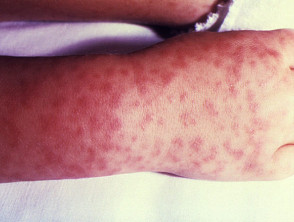
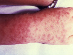
RMSF is classified as a moderate to severe infection, initially manifesting with an extended maculopapular rash Isolated or that evolves into petechial y petechial and purpuric, lesions, frequently presenting areas of necrosis. necrosis. Common systemic symptoms include abdominal pain and intense myalgia myalgia. Severe complications of RMSF may require multisystem intervention, including risk of encephalitis, encephalitis, renal ulceration, failure, hearing loss, and nerve nerves, damage, in addition to possible amputations secondary to necrosis. There is a significant mortality risk, which can reach up to 30% if antibiotic treatment is not administered promptly. mortality mortality.
African Tick-Bite Typhus
African tick-bite typhus is caused by Rickettsia africae and is predominantly found in sub-Saharan Africa and the West Indies. Livestock acts as the main host for ticks of the genus Amblyomma spp. carrying the infection. This condition tends to be more frequent during the period from November to April.
Clinically, African tick-bite typhus is usually asymptomatic asymptomatic or presents as mild severity, characterized by a maculopapular or vesicular. vesicular rash. A distinctive feature is that its eschar is often multiple, and it is common to observe groups of people affected simultaneously in shared exposure settings.
Complications associated with African tick-borne typhus include prolonged fever and arthritis. arthritis-like reactions. Fortunately, the mortality risk associated with this variant of rickettsiosis is very low compared to other severe forms of the disease.
Boutonneuse Fever
Boutonneuse fever, also called Mediterranean spotted fever, is caused by Caused by. Rickettsia conorii. It is transmitted mainly through Rhipicephalus sanguineus Rhipicephalus sanguineus ticks that parasitize rodents and dogs as primary hosts. Its distribution covers Southern Europe, Africa, Southern Russia, and Southern Asia, with higher prevalence between May and September.
Boutonneuse fever generally presents with mild severity. It begins with a macular macular rash.
Rickettsialpox
that rapidly evolves to maculopapular. Compared to other rickettsioses, the mortality risk associated with boutonneuse fever is low when adequate medical care is provided. Rickettsia akari Rickettsialpox (also known as Kew Gardens spotted fever or vesicular rickettsiosis) is caused by, Rickettsia akari, and is transmitted by the house mouse mite,.
Liponyssoides sanguineus.
, which infects rodents. This disease is detected in the United States, South Africa, the Balkan states, and Ukraine, predominantly affecting urban environments. It commonly arises after deratization, when mites, seeking a new blood source, bite humans. Rickettsia, Rickettsialpox usually manifests benignly, with the initial lesion (the papule that becomes a vesicle and involutes into an eschar) being a key feature for the differential diagnosis of spotted fevers. It is vital to differentiate it from RMSF due to implications in therapeutic management.
In summary, although all these infections belong to the genus papulovesicular. , the variety of the vector (tick, mite) and the causative bacterial species determine the clinical severity, rash pattern, and geographic distribution. Recognizing these distinctions is fundamental for implementing the correct antibiotic treatment and reducing morbidity associated with exanthematous rickettsioses. photophobia. may manifest asymptomatically or present with mild symptoms, generally characterized by a sparse.
papulovesicular rash. Despite its generally mild nature, central nervous system involvement has been documented, including symptoms such as neck stiffness and
photophobia. Rickettsialpox maintains a very low associated mortality risk. Flea-Borne Spotted Fever (Rickettsia felis). Flea-borne spotted fever is caused by the bacterium, Rickettsia felis. Its natural transmission cycle involves animal hosts such as cats, dogs, and rodents, spreading through the bite of the cat flea,, Ctenocephalides felis.
, globally. generalized. This pathology is generally classified as a mild disease, typically manifesting as a generalized maculopapular.
rash. Its peculiarities lie in being an emerging disease; it has only been identified in the last two decades, and its cases currently remain poorly defined and consequently underestimated in epidemiological statistics.
Although the mortality risk is low, its recent emergence and current underreporting underscore the need for constant surveillance to establish its true epidemiology and full spectrum of severity.
Spotted Fevers by Rickettsias: Clinical Identification, Diagnosis, and Therapeutic Approach.
Flea-borne spotted fever represents a diagnosis perhaps less recognized than the actual spectrum of *Rickettsia* diseases suggests. It is essential to understand the spotted fevers caused by these bacteria.
Consistent Clinical Characteristics of Rickettsial Spotted Fevers
- There is a fundamental symptomatic triad that accompanies most diseases transmitted by bacteria of the genus *Rickettsia*, characterized by:.
- High and intense fever.
- Severe headache, manifested as a throbbing pain.
Generalized myalgia, meaning widespread muscle pain.
- Evolution and Characteristics of the Skin Rash.
- The skin rash, the hallmark of these fevers, usually emerges between three and six days following the onset of febrile symptoms.
- Its development pattern typically begins on the patient's extremities and progressively spreads toward the trunk, frequently also affecting the palms of the hands and the soles of the feet.
- The presence of which facilitates clear visualization of the The exanthem is usually maculopapular in nature, although in certain occasions it may progress to a vesicular phase.
- The presence of petechiae is possible, indicating subcutaneous microhemorrhages that, in more severe scenarios, can evolve into areas of tissue necrosis. inoculation resection.
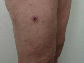
It is common to observe a cutaneous eschar—a characteristic dark scab—at the primary inoculation site, whether from a tick or mite bite, typically manifesting between 4 and 10 days after the initial inoculation.
Severe Complications Associated with Rickettsial Spotted Fevers
- Although many *Rickettsia* spotted fevers present a mild course, when the disease worsens, significant systemic complications may manifest:.
- Intense and persistent abdominal pain.
- Inflammation Development of tissue necrosis, a state that in extreme cases could warrant limb amputation.
- Orthostatic Severe pulmonary inflammation (pneumonitis).
- Diagnosis of anemia Severe hypotension, indicative of circulatory compromise..
- Diagnosis of acute renal failure.
- Rapid development of acute respiratory failure. Potential neurological damage.
- Circulatory collapse (shock) and subsequent multi-organ failure.
- Encephalitis, which involves direct inflammation of the brain tissue.
Key Steps in the Diagnostic Process for Spotted Fevers
The initial diagnosis of pathologies encompassed in the *Rickettsia* spotted fever group fundamentally depends on the clinical presentation observed in the patient and the accurate collection of their exposure history.
to environmental exposure to ticks or mites. Definitive laboratory confirmation is usually an intricate process that may require the implementation of several diagnostic techniques:
- Serology Serology (focused on detecting specific antibodies).
- Polymerase chain reaction (PCRPolymerase chain reaction (PCR).
- Cell culture. , used to identify the genetic material of the causative pathogen.
Cell culture, although this method is less frequent in daily clinical routine.
Critical Differential Diagnosis for Macular Fever
- The diagnostic approach must be broad, considering any pathology that manifests with the characteristic combination of fever, headache, and skin rash (exanthem). Priority conditions to rule out include:.
- Typhoid fever.
- Dengue. Typhoid fever. Rickettsiosis caused by.
- *Rickettsia* typhi.
- (known as murine typhus).
- Scrub typhus.
- Melioidosis.
- Various viral etiology infections.
- Measles and Rubella. hemorrhagic Secondary syphilis.
Other
- *Rickettsia* typhi.
- Tularemia.
- fevers.
- of viral origin.
- Furthermore, if the predominant clinical finding is the presence of an eschar (skin lesion), it is imperative to broaden the differential diagnosis to include:.
Scrub typhus.
Tularemia.
Anthrax (carbuncle).
Venomous spider bite. Unlike other Any other skin lesion of traumatic origin.
- Effective Antibiotic Treatment Against Spotted Fevers.
- Fortunately, rickettsioses, including spotted fevers, respond favorably to early antibiotic administration. Doxycycline, as a member of the tetracyclines, is established as the preferred first-line therapy. Chloramphenicol is strictly reserved as a second-line alternative treatment, especially indicated if allergies to the main drug or absolute medical contraindications for its use exist.
- Fundamental Prevention Strategies for Rickettsial Infections.
- Currently, no effective vaccine has been developed to proactively prevent.
rickettsial infections. Therefore, all preventive strategies focus rigorously on controlling and mitigating exposure to the main arthropod vectors: ticks and mites. Essential prophylactic measures include:
Constant and strict application of approved insect repellents, such as *N, N-diethyl-3-methylbenzamide* (DEET). sequelae Correct use of protective clothing when moving through areas with high tick density (this involves tucking pants into socks or wearing closed footwear). paralysis Thorough and periodic inspection of domestic pets to locate and remove any ticks.
Implementation of effective programs to control rodent populations, as they function as important infection reservoirs.


