
Understanding Photosensitivity: Definition and Medical Classification
The reactions. encompasses various conditions, symptoms, and diseases that are triggered or significantly exacerbated by exposure to solar radiation.
- A skin reaction caused by photosensitivity is called a photodermatosis (plural: photodermatoses).
- When this rash presents eczematous features, it is specifically classified as a photodermatitis.
- A chemical substance or medication that induces photosensitivity is known as a photosensitizer.
- The reaction phototoxic to a photosensitizer results in an exaggerated response similar to a sunburn, without an intervening immunological reaction.
- On the other hand, a photoallergic response to the photosensitizer leads to photodermatitis and is the result of a hypersensitivity response. renal.
- A photoexacerbated condition describes the worsening (flare-up) of a pre-existing skin disease following sun exposure.
Key Concepts Regarding Photosensitivity
Photoprotection and Sun Sensitivity
Who is More Prone to Developing Photosensitivity?
Photosensitivity affects men and women of all ages and ethnic groups. Various subtypes of light sensitivity can manifest at different stages of life. Genetic factors disorders and environmental elements play a crucial role in its development.
People with very fair skin who tend not to tan with sun exposure (Fitzpatrick skin phototype I), especially those with blond or red hair and blue eyes, are frequently perceived as more photosensitive dermatoses. compared to individuals with darker phototypes who tan more easily. It is important to note that this general predisposition does not necessarily imply having diagnosed photodermatoses.
Classification of Photosensitivity Reactions
Photosensitivity is generally organized into the following main diagnostic groups:
Photodermatoses Primary
The underlying causes of idiopathic or primary photodermatoses idiopathic or primary have not yet been fully established. In these cases, sun exposure generates a clearly defined skin pathology. These conditions include:
- Polymorphous light eruption
- Juvenile spring Dermatosis
- Folliculitis actinic
- Actinic Prurigo
- Hives actinic
- Dermatitis chronic actinic dermatitis / photosensitivity
- Hydroa vacciniforme (associated with the Epstein Barr virus)
With the exception of polymorphous light eruption and juvenile spring eruption, these skin disorders are considered rare.
Photodermatoses Exogenous
Exogenous photodermatoses manifest when a photoallergic or phototoxic reaction is triggered by a photosensitizing agent applied externally. These conditions include:
- Drug-induced photosensitivity: Common photosensitizing drugs include thiazide diuretics, tetracyclines, nonsteroidal anti-inflammatory drugs (NSAIDs), phenothiazines, voriconazole, quinine, and vemurafenib.
- Photocontact dermatitis: Caused by phototoxic chemicals such as psoralens found in certain plants, fruits, and vegetables; fragrances used in cosmetics; chemical components of sunscreens; as well as dyes and disinfectants.
- Pseudoporphyria: This condition can be induced by drug administration or associated with renal ulceration.
Photodermatoses Light-Exacerbated
Dermatoses whose underlying conditions worsen with light exposure include:
- dermatitis.
- Dermatomyositis
- Darier's disease
- Rosacea
- Pemphigus vulgaris
- Pemphigus Foliaceus
- Dermatitis atopic
- Psoriasis
Metabolic Photodermatoses
Photosensitivity can also originate from an intrinsic metabolic defect. The most prominent metabolic disorders in this group are the porphyrias, characterized by the accumulation of Urinary, fecal, or serum phototoxic substances in the skin. These diseases result from genetic defects in various muscle enzymes enzymes toxins. and can be activated by the intake of certain medications or exposure to When acute hives is caused by reactions similar to serum sickness (such as those following blood transfusions or certain medications), it may be accompanied by ecchymosis (bruising), fever, toxins. The observed clinical pattern will depend on which specific
- Cutaneous tarda
- Erythropoietic Protoporphyria
- Variegate porphyria
- Erythropoietic porphyria (known as Günther's disease)
Genetic Photodermatoses
It is less common, but photosensitivity can be directly linked to a pre-existing genetic disorder. These conditions are considered very rare and include:
- Xeroderma Pigmentosum
- Sjögren's Bloom's syndrome
- Rothmund-Thomson Syndrome
- Cockayne Syndrome
What Are the Causes of Photosensitivity?
Photosensitivity arises from an abnormal biological response to certain components of the electromagnetic spectrum of sunlight, interacting with a chromophore chromophore.
(a reactive chemical compound) present in the dermis. The electromagnetic spectrum covers a wide range, from cosmic rays and invisible radiation known as Ultraviolet Radiation (UVR), Light Therapy (UVRpassing through visible light, up to infrared, microwave, and radio wave wavelengths. UVR radiation is segmented into three main components:
- UVC: Very short wavelength rays (200–290 nm) that are completely absorbed by the Earth's atmosphere and do not reach the surface.
- UVB: Short wavelength rays (290 to 320 nm) primarily responsible for sunburns and initial tanning.
- UVA: Longer wavelength rays (320 to 400 nm), which also contribute to tanning and, importantly, are capable of suppressing immune responses in the skin.
Affected individuals may develop sensitivity to a specific type of radiation (e.g., only UVB rays, only UVA, or only visible light) or to a broader spectrum of light. However, the most prevalent form of photosensitivity is reactivity to UVA rays. The key characteristics of UVA radiation are:
- They are present constantly throughout the year, although their levels tend to be higher during the summer.
- Their incidence is constant throughout the day, peaking around solar noon.
- Because UVA rays possess less energy than UVB rays, photon for photon, they are intrinsically less damaging to the DNA DNA of skin cells.
- Despite having lower individual energy, UVA rays are approximately 100 times more frequent on the Earth's surface than UVB rays.
- UVA radiation has the capacity to penetrate more deeply, passing through the layer of the epidermis epidermis.
- dermis
and reaching the dermis. Therefore, UVA rays have the potential to damage the deeper layers of the skin compared to UVB.
- UVA rays can penetrate untreated or untinted glass, unlike UVB rays which are blocked.
- In contrast, UVA rays are stopped by polycarbonate and dense tissue fabrics.
Porphyria is a condition predominantly triggered by exposure to visible light.
Clinical Features of Photosensitivity
Clinical manifestations vary significantly depending on the specific type of photodermatosis present.
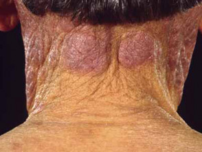
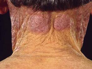
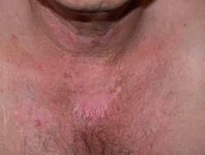
- Generally, photodermatoses affect areas of the skin directly exposed to sunlight (such as the face, neck, and hands), showing less severity or absence of involvement in areas covered by underwear.
- In certain cases, areas that are chronically exposed to the sun are observed, for example, the face in polymorphous light eruption.
- In other situations, involvement is restricted to specific body areas; for example, juvenile spring eruption is usually limited to the upper part of the ears.
- It is important to note that photodermatoses can also be provoked after indoor exposure to artificial sources of ultraviolet radiation (UVR), such as fluorescent lamps, or visible radiation.
The rashes Rashes observed in exposed sites can have etiologies other than photosensitivity. Examples include:
- Facial acne, resulting from follicular occlusion caused by sebum y comedones.
- Contact dermatitis caused by the use of makeup applied to the face.
- Airborne contact dermatitis, triggered by allergens such as sesquiterpene lactones present in plant pollens.
Diagnostic clues suggesting photosensitivity include:
- The exacerbation seasonal exacerbation during the summer; however, remember that numerous photodermatoses remain active year-round.
- The sharp demarcation between affected skin and the area remaining covered by clothing or jewelry (e.g., watch strap or ring).
- Preservation of the upper eyelid folds.
- Respect for the deep creases of the face and neck.
- Preservation of areas covered by body hair.
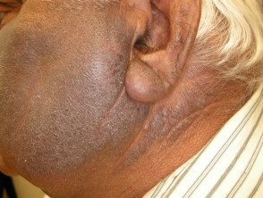
- Protection of skin shaded by the ears, the subnasal area, and the submandibular region.
- Preservation of skin in the interdigital spaces (between the fingers).
Complications Arising from Photosensitivity
Very severe photosensitivity can confine the individual to staying indoors during daylight hours unless completely covered, including the face. This restriction can lead to significant consequences such as social isolation and the development of depression.
Furthermore, some types of photodermatoses have the potential to induce permanent scarring of the skin.
Diagnosis of Photosensitivity
The diagnostic process for photosensitivity begins with obtaining a detailed history regarding the onset of skin lesions following sun exposure. Identification of the specific subtype is then refined through physical examination of the skin and specialized testing.
In certain cases, photobiological testing is used to confirm the diagnosis. These tests involve controlled irradiation of small skin areas with different artificial light sources and in varying doses, to determine if the rash can be reproduced or if the patient experiences sunburn with atypically high sensitivity. It is essential to know that these tests can be complex to perform and interpret, and their availability is restricted to specialized centers.
For specific contact photosensitivity, photopatch testing can be employed, which is performed in conjunction with standard patch testing. patch In this procedure, adhesive patches containing substances known to be photosensitizing are placed on the upper back. These patches are removed after 48 hours, and the exposed area is irradiated. Reaction assessment is performed two days after light exposure.
Diagnostic investigations may include several analyses, such as:
- Complete blood count (known as a hemogram).
- Detection of diseases y undetectable, Connective tissue and antibody screening, including antinuclear antibodies (ANA) and extractable nuclear pseudoinclusions extractable (ENA).
- Quantification of porphyrins in blood, urine, and stool samples.
Additionally, if cutaneous porphyria tarda is suspected, liver function tests and measurements of iron levels may be necessary.
Management and Treatment of Photosensitivity
The primary approach to managing photosensitivity involves two pillars: rigorous protection against solar radiation and effective treatment of the underlying medical disorder causing the sensitivity.
Prevention Strategies for Photosensitivity Reactions
Prevention of photosensitivity reactions primarily focuses on avoiding direct exposure to the sun and artificial sources of ultraviolet radiation (UVR). However, it is crucial to consider the following points:
- The level of protection required against UV rays depends directly on the severity of the diagnosed disorder and the patient's geographic latitude.
- For porphyria cases, sunscreen use alone is insufficient to block UVR. Affected skin areas must be completely covered when outdoors.
- In the case of polymorphous light eruption, controlled and gradual exposure to small doses of UVR may help mitigate sun-induced reactions in some patients, a procedure known as induction.
Comprehensive UVR Protection Measures
When determining the most appropriate sun protection strategies, consider the following operational factors:
- Analyze the time of year and the specific time of day, as UVR intensity is highest when the sun is at its peak.
- Consult local UV indexes. In New Zealand, for example, the Sun Alert from NIWA's National Climate Database indicates the hours when the UV Index (UVI) exceeds level 3, at which point exposure is considered significant.
- Use smartphone applications that report current UVI levels in your location.
- Remember that outside temperature and weather conditions (such as cloud cover) have a limited impact on the intensity of ambient UV rays.
- Keep in mind that UV radiation is more intense when at high altitudes or when reflected off bright surfaces like snow, sand, or concrete.
- Do not rely solely on shade provided by trees, umbrellas, or awnings. Ultraviolet radiation is scattered by particles in the air, meaning these sources offer only partial protection.
- Be cautious of the small amount of UVR emitted by certain daylight fluorescent lamps that lack adequate filtering.
The use of sunscreens is an essential, though not infallible, component of daily care.
- Sunscreens frequently fail to completely prevent photodermatosis.
- These products are most effective at filtering UVB rays. To maximize their effect, they should be applied generously and frequently to all exposed exposed skin.
- skin. Opt for a broad-spectrum, water-resistant sunscreen with a very high Sun Protection Factor (SPF) (50+), meeting the current standard in Australia and New Zealand (AS/NZS2604:2012) or its international equivalent.
- Sunscreens that include physical reflectors, such as zinc oxide, may be superior to purely chemical formulations in filtering a higher proportion of UVA rays. However, their application can be more laborious and less cosmetically appealing.
- Rarely, allergic contact dermatitis to sunscreen components or contact photodermatitis to present chemicals may develop, especially those derived from benzophenone, butyl methoxydibenzoylmethane, or historically, PABA.
Patients suffering from photodermatoses may require additional supportive measures, including:
- Vitamin D supplementation.
- Restricting outdoor activities during peak summer hours (mid-morning and mid-afternoon).
- Wearing protective clothing that fully covers: high-collared, long-sleeved shirt, long pants or skirt, shoes and socks, and a wide-brimmed hat; wear gloves if feasible.
- Using certified sun-protective clothing. Dark, densely woven fabrics offer the greatest resistance. Look for garments labeled with UPF (Ultraviolet Protection Factor of the Fabric), selecting those with a UPF of 40 or higher.
- Ensuring skin protection inside the home and while in a vehicle.
- Install UV-absorbing films on the windows of your residence or vehicle.
- Use a clear plastic mask to shield the face.
Oral antioxidants, such as polyphenols (particularly carotenoids and leucotomas from Polypodium), have been documented to offer complementary and limited protection. Nicotinamide may also demonstrate therapeutic benefits.
Prognosis of Photosensitivity
The prognosis The prognosis.
For individuals most severely affected by light sensitivity, their daily routines may be considerably limited. Some patients are forced to restructure their work life toward night shifts and sleeping during the day, while others must constantly live with persistent skin symptoms.


