
Pseudoxanthoma Elasticum (PXE): Fundamentals, Key Histology, and Diagnostic Methods
Pseudoxanthoma elasticum (PXE) is defined as a rare connective tissue disorder diseases, characterized by the fragmentation followed by the mineralization of elastic fibers. elastic fibers. These structural alterations primarily impact the skin, vision (eyes), and the cardiovascular system. The condition is genetically linked to more than 110 mutations distinct mutations identified in the gene ABCC6 gene.
Histological Analysis of Pseudoxanthoma Elasticum
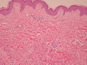
From a histopathological perspective in cases of PXE, the elastic fibers located in the reticular dermis dermis reticular show an evident pattern of fragmentation. These abnormal structures exhibit strong basophilic staining can be identified and are easily observable in sections stained with Hematoxylin and Eosin (H&E). In certain instances, the presence of these altered fibers causes a granulomatous response granulomatous (as illustrated in Figures 1, 2, and 3).
Molecular Pathology in Pseudoxanthoma Elasticum
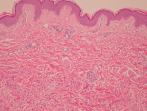
Figure 1
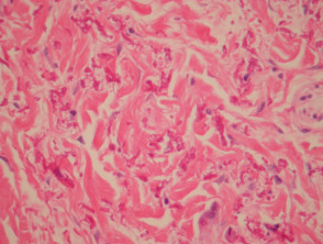
Figure 2
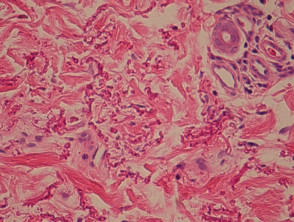
Figure 4
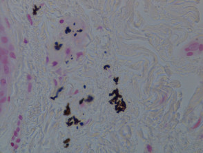
Figure 5
Special Studies for Diagnostic Confirmation of PXE
Von Kossa staining is crucial in the diagnostic process, as it clearly visualizes the altered elastic fibers, which are observed coated with calcium deposits (a finding highlighted in Figure 4). This staining technique unequivocally highlights the pathognomonic **calcification** of the disease.
Differential Diagnosis Determination in Pseudoxanthoma Elasticum
Establishing the differential diagnosis requires a meticulous examination, especially when contrasting with the appearance of healthy skin. Without an exhaustive, high-magnification analysis of the reticular dermis, reticular dermis, , abnormalities S100 protein. elastic fiber. the abnormalities in the elastic fiber can be easily overlooked.
It is essential to directly correlate histopathological findings with observed clinical manifestations, as elastic fibers with morphological characteristics similar to PXE can appear sporadically in various dermal pathologies. dermal.
A condition that can clinically resemble PXE is Papular Dermal Elastolysis (which causes the appearance of fibroelastic papules). papules However, histologically, this entity is differentiated by presenting a decrease or even the total absence of elastic fibers in the papillary dermis. papillary dermis.
To achieve a definitive diagnosis of pseudoxanthoma elasticum, precise integration of clinical findings, available genetic data, and a detailed histopathological study using specific stains that allow for certain distinction from other associated skin diseases is required.


