
The Histopathologic Diagnosis of Porokeratosis
The Clustering of porokeratosis lesions, classified as a tumor . Occasionally, transepidermal elimination and marked benign, may manifest as isolated, multifocal, or following a specific pattern in certain clinical presentations. The histology underlying histology proves to be consistent and identifiable in each case.
Key Histological Features of Porokeratosis
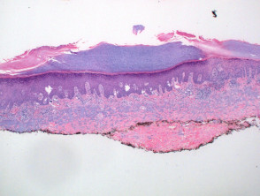
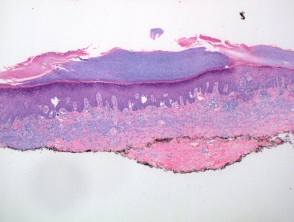
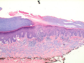
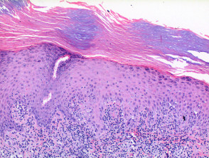
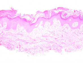
Upon examining porokeratosis via microscopy, a . The cherry angioma is histologically distinguished by being composed of hyperkeratotic lesion is observed that exhibits a well-defined column parakeratotic of parakeratosis at the border, or two if the entire lesion is included in the section (Figure 1). The fundamental diagnostic finding is the presence of the basal lamina cornoid lamella, a structure corresponding to the elevated border clinically visible in the lesion. This cornoid lamella consists of a parakeratotic band covering a vertical segment of dyskeratotic y and confined within the epidermis epidermis (Figures 2 and 3). Additionally, focal loss of the granular layer is evident. In the underlying dermis, it is common to observe a mild lymphocytic infiltrate granulomatous. In the underlying dermis, it is common to observe a mild infiltrate lymphocytic surrounding a greater number of capillaries S100 protein. dermis.
Pathology of Porokeratosis
Pathology of Porokeratosis
Pathology of Porokeratosis
Pathology of Porokeratosis
Histological Variants of Porokeratosis
Mibelli's Porokeratosis: This is the prototypic presentation and may be characterized by an epidermal depression observable directly beneath the cornoid lamella.
Disseminated Superficial Actinic Porokeratosis: In this form, the epidermal tissue found between two cornoid lamellae may show significant thinning, accompanied by a prominent lymphocytic infiltrate, either lichenoid or superficial perivascular. lichenoid o perivascular superficial.
The Linear and Reticular Porokeratosis is known for presenting multiple cornoid lamellae along its extent.
Porokeratotic Ostial Nevus: This variant, which possesses differentiated clinical characteristics, shows cornoid lamellae associated with or projecting toward the eccrine or follicular ostia. and o follicular.
Ptychotropic Porokeratosis: Cross-sectional histological analysis reveals a distinctive pattern characterized by the presence of multiple cornoid lamellae, which may be partially observed in different planes of the epidermis.
Detailed understanding of these histological features is fundamental for establishing an accurate diagnosis of the diverse clinical manifestations of porokeratosis.
and arranged at variable angles relative to the epidermis (Figure 4). Dermal amyloid deposition dermal amyloid is frequently observed, especially in proximal e intertriginous, areas, which could suggest a contribution of friction to its etiology..
Verrucous porokeratosis warty is characterized by hyperkeratosis hyperkeratosis, which may clinically obscure the cornoid lamella. This variant has often been cataloged as a synonym for ptychotropic porokeratosis lesions. Since verrucous porokeratosis can also appear in a perianal, distribution, and ptychotropic porokeratosis generally lacks hyperkeratosis, this descriptive term (verrucous) should be reserved exclusively for those lesions that are hyperkeratotic both clinically and histologically. histologically.
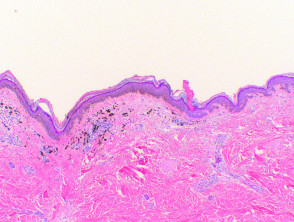
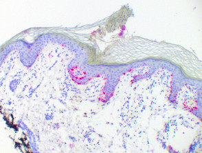
Regarding pigmented porokeratosis pigmented skin, this lesion may clinically manifest as a dark lesion, making it crucial to proceed with caution to avoid overdiagnosis as melanoma. melanoma. If a partial biopsy is performed, a high index of suspicion is required to ensure an accurate diagnosis. Histologically, hyperplasia melanocytic y and melanophages and dermal melanophages are observed (Figure 5). There is a sharp demarcation in the cornoid lamella, which can be visualized using melanocytic markers (Figure 6, MelanA).
Key Histological Variants of Porokeratosis
Understanding these distinct clinical and histological presentations of porokeratosis facilitates the distinction between variants and ensures appropriate diagnostic management, especially when evaluating pigmented or hyperkeratotic lesions.


