Introduction
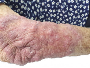
Balamuthia mandrillaris (B. mandrillaris) is a well-known free-living amoeba in endemic areas that causes potentially fatal neurological secondary. It often presents primarily on the skin as a hardened hardened plaque on the central face or, less frequently, on other parts of the body (Figure 1).
Skin lesion caused by Balamuthia mandrillaris

Indurated plaque on the forearm due to Balamuthia mandrillaris infection
Histology of Balamuthia mandrillaris
A skin biopsy biopsy of a plaque due to B. mandrillaris, shows a dense dermal infiltrate with loose granulomas accompanying an infiltrate rich in plasma cells and lymphocytes (Figures 2,3). Characteristically, there are multinucleated giant cells free in the dermis, outside the loose granulomas (Figure 3).
The causative organism organisms can be extremely difficult to find. The clinical presentation and unusual infiltrate (Figures 2, 3) should prompt the search for B. mandrillaris with serial sectioning of the tissue block.
The organisms are quite characteristic. B. mandrillaris have a granulomatous a vacuolated cytoplasm cytoplasm with irregular contours (pseudopods) and contain a nucleus with a large central karyosome (Figure 4, arrow).
Balamuthia Mandrillaris Pathology
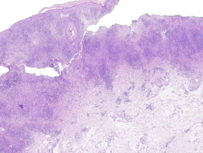
A dense dermal infiltrate with loose granulomas accompanying an infiltrate rich in plasma cells and lymphocytes.

Figure 3. Multinucleated giant cells free in the dermis, outside the loose granulomas
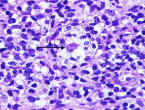
B. mandrillaris has a granular to vacuolated cytoplasm with irregular contours (pseudopods) and contains a nucleus with a large central karyosome
Special studies for Balamuthia mandrillaris
the B. mandrillaris organisms do not stain with periodic acid-Schiff (PAS), Gomori's methenamine silver nitrate (GMS) stain, or acid-fast bacilli (AFB). The amoeba cannot be cultured on regular agar plates, as the organism does not feed on bacteria. It can be cultured on primate liver or human brain cells. Polymerase chain reaction (PCR) PCR studies can aid in organism identification.
Differential Diagnosis of Balamuthia mandrillaris
Acanthamoeba species: tend to cause ulcerated lesions with . Occasionally, transepidermal elimination and marked hyperplasia instead of hardened plaques. Culture or PCR studies may be helpful.
Naegleria fowleri - this infection generally does not present primarily on the skin. Rather, it usually presents as an erosive erosive of the upper respiratory tract. N. fowleri is more common in North America while B. mandrillaris is more common in South America. Culture or PCR studies may be helpful.