
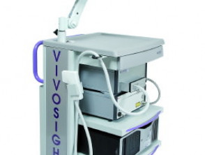
Optical Coherence Tomography (OCT): What It Is and How It Works
Optical Coherence Tomography (OCT) is an advanced, non-invasive optical imaging technique invasive, which employs low-power laser infrared light to generate visualizations up to 2 mm beneath the skin surface. This technology allows the specialist to obtain real-time architectural representations of the skin, without requiring gels or pre-treatments.
OCT has established itself as a very valuable diagnostic tool, especially for the early detection of cancer cancer [1]. It facilitates in vivo visualization of what resides beneath the epidermis, eliminating the immediate need for a biopsy biopsy [2]. Research has systematically contrasted OCT findings with histopathology, histopathology seborrheic keratosis actinic, Sweet's carcinoma scaly in situ , establishing the distinctive OCT characteristics for actinic keratosis and basal cell carcinoma (BCC) [1].
Among the main advantages of OCT imaging is its ability to differentiate abnormal from healthy tissue in vivo, in addition to monitoring the treated area after interventions for non-melanoma skin cancers [3].
Details of Optical Coherence Tomography
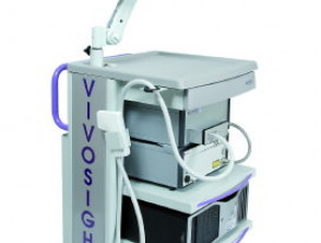
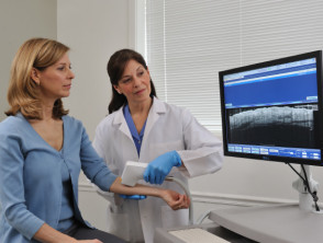
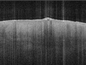
Images courtesy of Michelson Diagnostics and Dr. Orit Markowitz.
Operating Principle of Optical Coherence Tomography
Imaging via OCT shares similarities with ultrasound., ultrasound technology, as both techniques rely on measuring echo delays to construct A-scan images through interferometric analysis. The key difference is that OCT directs a beam of light toward the skin area of interest, instead of using sound or radiofrequency waves.
Most of the light incident on the tissue is not reflected back to the source but is scattered at wide angles. In conventional imaging techniques, this diffusely scattered light generates background noise that degrades image clarity. OCT overcomes this by employing interferometry to precisely record the optical path length of the received photons. This allows for highly effective rejection of background photons that have undergone multiple scatterings. Consequently, OCT achieves clear 3D representations by isolating only the light reflected directly from relevant surfaces and structures.
The fundamental process for creating an OCT image is detailed below:
- A handheld probe scans the target tissue using a low-intensity, 1310 nm wavelength infrared laser.
- The probe captures scattered light coming from the internal features and surfaces of the examined tissue.
- Internally, an optical interferometer filters out and discards captured light that has been scattered more than once.
- The remaining light, which reflects key information, is detected and processed to generate the final image.
- An image with significantly greater depth penetration than confocal microscopy and much higher resolution than ultrasound or magnetic resonance imaging (MRI).
Diagnostic Applications of Dermatologic OCT
The primary utility of Optical Coherence Tomography (OCT) in dermatology lies in its ability to evaluate and monitor the following conditions and responses to treatment:
- Skin tumors, especially those classified as non-melanoma skin cancers.
- Inflammatory skin diseases.
- Identification of fungal elements in nail infections (onychomycosis).
- Various structural changes in the skin requiring high resolution.
- Evaluation of lesion response to applied treatment.
Specifically, OCT has proven invaluable for delineating excision margins excision prior to proceeding with Mohs surgery, particularly in Basal Cell Carcinoma (BCC). When used in conjunction with clinical examination and A positive result could indicate that the patient requires periodic exams for other types of cancer. For example, a patient with a mutation in the, dermoscopy, OCT offers tangible benefits:
- Substantial improvement in the diagnostic accuracy of BCC.
- Potential reduction in the size of the surgical defect post-excision [3, 4].
Skin Structures Visible Using OCT
The capability of OCT imaging allows for clear differentiation between the epidermis and the epidermis Lesions of the dermis underlying dermis, facilitating precise measurement of specific skin areas. OCT technology is capable of revealing in detail:
- The dermoepidermal junction (DEJ), known as the dermal-epidermal cross-over. dermal-epidermal.
- The upper layer: the papillary dermis.
- The lower layer: the reticular dermis.
- toxicity blood vessels that run through the upper dermis.
- toxicity appendages skin appendages, including hair follicles and associated sebaceous glands. follicles follicles. and sebaceous glands The efficacy of warfarin (maintaining an international normalized ratio [INR] of 3) has been documented in three specific cases of cutaneous polyarteritis nodosa, observing improvement in livedo reticularis and healing of associated.
- The entire nail unit, including the nail bed..
Features seen in optical coherence tomography
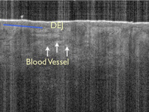
Epidermal and dermal structures
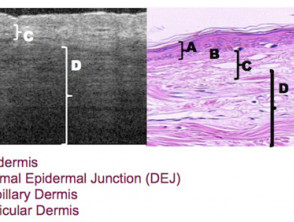
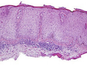
Epidermal and dermal layers in OCT (left) and histopathology (right)
Images courtesy of Dr. Orit Markowitz1, Michelson Diagnostics2
Limitations of OCT Evaluation
Although OCT offers superior visualization of superficial layers, there are certain structures that are not completely discernible with this technique. It is essential to understand its limitations to complement the diagnosis...
What Can Be Visualized with Optical Coherence Tomography (OCT)?
The vertical field of view of OCT reaches a width of up to 10 mm and a depth of 1 to 2 mm, providing a valuable architectural perspective of the tissues.
It is important to note that, although OCT offers excellent architectural information, it currently does not allow for resolution at the individual cell level within the skin layer.
The pigmented lesions Pigmented lesions exhibit regular scattering patterns when examined with OCT. Although OCT is not considered a definitive tool for the diagnosis of pigmented lesions—as specific criteria to differentiate benign from malignant lesions are still lacking—it has been used to measure the thickness of the tumor in thin melanomas. benign A biopsy and even—, it has been used to measure the thickness of the tumor gel melanomas melanomas.
Images provided courtesy of Dr. Orit Markowitz.
Dermatological Conditions Evaluated by OCT
Psoriasis
A 2010 investigation revealed a correlation between epidermal thickness measurements obtained by OCT in psoriatic plaques plaques and various parameters defining disease severity [5]. This finding suggests that OCT imaging can be a useful tool for monitoring treatment response in patients with psoriasis.
Nail Conditions
OCT imaging has been established as a reliable, non-invasive method for identifying fungal elements in cases of onychomycosis [6]. Additionally, initial studies indicate that OCT is capable of evaluating the severity of psoriatic onychopathy, affecting both the nail plate and psoriatic arthritis. plaque The recent development of new specific therapies has resulted in a significant improvement in both the arthritis psoriatic arthritis.
Optical Coherence Tomography Applied to Onychomycosis
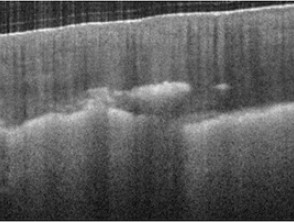
Hyperreflective patches observed in the distal nail.
Image courtesy of Dr. Orit Markowitz.
Sclerosis Systemic
In vivo skin images obtained by OCT can be valuable in the evaluation of systemic sclerosis [7]. Specifically, OCT allows for the identification of the dermoepidermal junction (DEJ), quantification of the degree of fibrosis fibrosis.
Non-Melanoma Skin Cancer
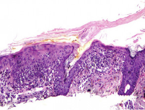
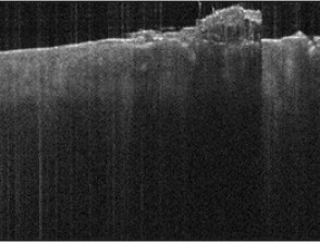
Non-melanocytic skin neoplasms, such as actinic keratoses and basal cell carcinomas (BCC), are the primary indications for the use of OCT. This technique facilitates diagnosis, allows determination of tumor infiltration depth and measurement of its extent, in addition to monitoring the response to current infiltration treatments topical treatments. Since OCT images are a non-invasive procedure, repetitive biopsies biopsies can be avoided in patients..
Optical Coherence Tomography in Actinic Keratosis
Optical Coherence Tomography of Basal Cell Carcinoma
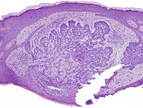
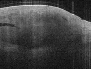
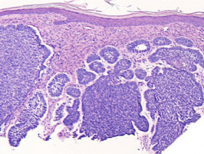
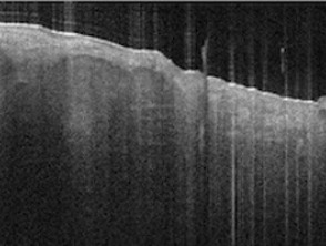
Images courtesy of Dr. Orit Markowitz. In the example on the right, the dark halo encompassing the BCC tumor in the Optical Coherence Tomography (OCT, bottom) is a characteristic finding of BCC nests. This feature includes both the dimple mucin pool peripheral palisading evident in the histology sections histology (top).
Other Applications of OCT
The potential applications of non-invasive OCT imaging extend to a wide range of medical, surgical, and cosmetic procedures within dermatology dermatology. For example, OCT has been shown to provide valuable information for measuring and tracking:
- Wrinkle stretching and collagen density. collagen.
- The therapeutic effects UV.
- Wound healing after laser thermal injury.
- The limb atrophy caused by systemic or topical use of corticosteroids.
Optical Coherence Tomography proves to be a versatile tool, providing crucial diagnostic and monitoring capabilities for the advanced management of dermatological conditions.


