
¿What is Neurofibromatosis?
Neurofibromatosis (NF) is a disorder genetic that affects the bone, soft soft tissue, tissue, skin, and nervous system. The clinical manifestations of this condition usually increase over time. Other Lesions Classified as Connective Tissue Nevi clinical manifestations of this condition usually increase over time.
There are at least 8 biopsy different clinical phenotypes of NF. Generally, it is classified into two main types:
- Neurofibromatosis Type 1 (NF1)
- Neurofibromatosis Type 2 (NF2).
Neurofibromatosis Type 1 (NF1)
NF1 affects approximately 1 in every 3,000 births and is also known as von Recklinghausen's disease. It is distinguished by the presence of:
- Six or more macules café au lait macules: flat, light brown spots on the skin.
- Freckling in the skin folds.
- Fatty Lisch nodules in the iris of the eye.
- Multiple neurofibromas: tumors that may protrude or hang from the skin.
Neurofibromatosis Type 2 (NF2)
NF2 occurs in about 1 in every 50,000 births. It is also known as bilateral acoustic neurofibromatosis or central neurofibromatosis. It is characterized by the development of multiple tumors and lesions lesions in the brain and spinal spinal cord.
It is important to note that a solitary neurofibroma neurofibroma is not associated with NF1 or NF2.
What Causes Neurofibromatosis?
Both NF1 and NF2 result from defects in different genes. genes.
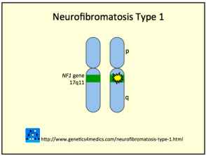
- NF1 is caused by a , originating from a in the gene neurofibromin gene mutation, located in the pericentromeric region of chromosome 17. chromosome 17.
- NF2 is due to a mutation in chromosome 22.
The mutated gene can be inherited from an affected parent through an autosomal autosomal dominant transmission, or it can arise as a founder gene due to a spontaneous mutation. If a parent has NF, there is a 50% chance of transmitting the gene to each of their children.
Genetics of Neurofibromatosis Type 1 and 2 *
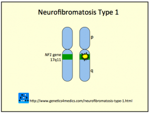
Neurofibromatosis Type 1
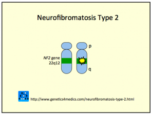
Neurofibromatosis Type 2
* Image courtesy of Genetics 4 Medics
What Are the Signs and Symptoms of Neurofibromatosis?
The extent and severity of the signs and symptoms of neurofibromatosis are highly variable among affected individuals. The specific clinical manifestation will depend on the type of NF (mainly NF1 or NF2) and the progression of the disease over time. Early detection of these indicators is essential for effectively managing the condition.
Characteristics and Manifestations of Neurofibromatosis
The severity of Neurofibromatosis (NF) manifestations varies significantly between individuals and can even differ within the same family.
Neurofibromatosis Type 1 (NF1)
Café au Lait Macules
Café au lait macules are well-defined, oval or irregular-shaped skin spots, characterized by a light brown color measuring more than 0.5 cm in diameter. These may be present from birth and their number tends to increase during the child's first years of life.
While it is possible to find isolated café au lait macules in many people who do not have NF, the presence of more than five of these spots considerably increases the probability of having NF1, especially if they appear during the first five years of life.
Statistically, more than five café au lait macules are observed in 1.8% of newborns, in 25-40% of diagnosed children, and in 14% of adults with NF1.
Axillary Freckling (Crowe's Sign)
Freckles located in the armpits are known as Crowe's sign and are a distinctive characteristic of Neurofibromatosis Type 1. These freckles typically emerge after the appearance of café au lait macules and before the development of neurofibromas, generally manifesting during puberty. They can also be observed in other skin folds, such as the groin.
Cutaneous Manifestations of Neurofibromatosis
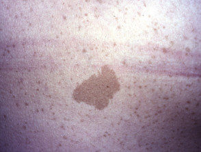
Café au lait mark
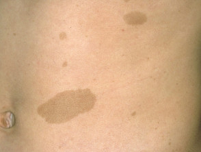
Café au lait mark
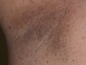
Axillary freckles
Lisch Nodules
Lisch nodules are small tumors that develop in the iris of the eye. After puberty, these nodules are present in 97% to 100% of patients with NF1. Although they usually do not cause functional problems clinically, their presence is crucial for confirming the diagnosis of the disease.
Neurofibromas
Neurofibromas are tumors composed of Schwann cells, fibroblasts, mast cells, and vascular components. They have the capacity to develop at any point along a nerve. They are classified into three main types: cutaneous, subcutaneous, and plexiform. While cutaneous and subcutaneous neurofibromas can present in other conditions, plexiform neurofibromas are exclusive to NF1.
- Cutaneous neurofibromas manifest as superficial, soft, well-circumscribed nodules, which can be brown, pink, or skin-toned, presenting a soft or firm consistency. It is possible to observe a pathognomonic indentation (dimpling) when pressing them with a finger. These do not present malignant potential.
- Subcutaneous neurofibromas are similar to cutaneous ones, but their origin is deeper. Occasionally, they may cause localized pain or tenderness.
- Plexiform neurofibromas appear as lobulated masses within the skin. Nodular plexiform neurofibromas affect the dorsal nerve roots, while diffuse plexiform neurofibromas are invasive tumors capable of affecting all layers of the skin, muscles, bones, and blood vessels.
More about Neurofibromatosis
The diagnosis and monitoring of Neurofibromatosis Type 1 manifestations are fundamental for clinical management, given the wide variability in symptom presentation throughout the patient's life.
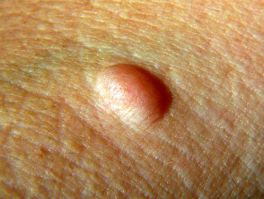
Neurofibroma
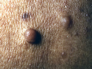
Neurofibromas
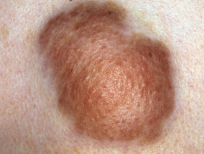
Plexiform neurofibroma
View more examples of the cutaneous manifestations of neurofibromatosis below.
Other Characteristics of Neurofibromatosis Type 1 (NF1)
The severity of cutaneous manifestations in NF1 does not always correlate directly with the extent of the underlying disease, as internal complications are common and often represent the greatest clinical challenge. Systemic involvements may include:
- Long bone malformation (especially below the knee and elbow) and development of spinal curvature (scoliosis).scoliosis).
- Short stature due to growth hormone deficiency or resistance.
- Learning difficulties (especially speech problems) and behavioral disorders; affects 25–40% of cases, with 5–10% experiencing some degree of intellectual disability.
- Development of tumors in the optic nerve (optic gliomas), which can lead to progressive vision loss.
- High blood pressure and other abnormalities in the blood system.
- Presence of tumors in the brain and spinal cord, which increases the risk of developing epilepsy.
- Lesions or tumors in the gastrointestinal tract that may cause obstruction or hemorrhage.
- Hearing impairment due to involvement of the auditory nervous system.
While most tumors associated with NF1 are benign benign (non-cancerous), it is estimated that people with NF1 have an elevated risk (between 3 and 15%) of developing or develop malignant tumors throughout their lives.
Neurofibromatosis Type 2 (NF2)
Unlike NF1, NF2 is characterized by fewer obvious external signs; it focuses primarily on the formation of multiple tumors and lesions within the brain and spinal cord. The most frequent initial symptom is usually bilateral or unilateral hearing loss caused by a symptom most frequent initial symptom is usually bilateral or unilateral hearing loss caused by a tumor tumor growing on one or both nerves vestibular nerves (auditory nerves). This manifestation often does not become evident until late adolescence or early twenties.
Tumors associated with NF2 are generally benign. However, the growth of these tumors within the skull and spine can compress vital structures, severely affecting essential bodily functions.
Clinical Follow-up of Patients with Neurofibromatosis
The fundamental goal of follow-up in patients with neurofibromatosis is the constant monitoring of tumor development and the implementation of early interventions when necessary.
- Children diagnosed with NF without symptoms should be examined and monitored every 6 to 12 months by their pediatrician.
- Those presenting with visual signs and symptoms require periodic evaluation and supervision by a specialized ophthalmologist specialist.
- Hearing deficiencies should be evaluated and followed up by an otolaryngologist (ENT).
- A , a pediatrician or a A neurologist must examine the patient if any neurological symptom or sign is present severe neurological, such as motor clumsiness, numbness, or weakness in the extremities.
To locate the site of any suspected tumor, advanced imaging studies can be scheduled, including ultrasound., ultrasound scans, computed tomography (CTImaging Tools for Diagnosis scans), and magnetic resonance imaging (MRI). or Magnetic Resonance Imaging (MRI).
Available Treatment Options for Neurofibromatosis
Currently, there is no definitive cure for neurofibromatosis.
Neurofibromas that grow significantly, cause pain, or present a risk of or evidence of lymphovascular invasion are key factors pointing towards malignancy or other complications may be surgically removed. Facial deformities can be addressed through cosmetic and reconstructive surgery.
Genetic counseling and exhaustive education about the disease are essential components of comprehensive management. A crucial consideration is the significant risk of social isolation or loneliness faced by people with NF. Future complications and the appearance of disfiguring lesions can cause these individuals to withdraw socially.
Molecularly targeted therapies show promising results as a future approach for the specific treatment of the various forms of neurofibromatosis.


