

Necrotizing Fasciitis: Histopathological and Bacteriological Diagnosis
The necrotizing fasciitis represents a critical medical emergency, classified as a rapidly progressive form progressive of destructive cellulitis. The most frequent etiologic agent involves Group A betahemolytic streptococci , although various other bacteria have also been associated with its development.
Histopathology of Necrotizing Fasciitis
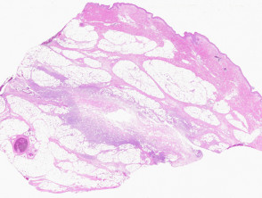
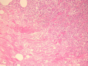
The definitive diagnosis of necrotizing fasciitis often requires obtaining a biopsy deep biopsy (see Figure 1). This histological analysis reveals an intense acute inflammatory reaction inflammatory acute primarily affecting the subcutaneous fatty tissue subcutaneously (Figure 2). It is important to note that this infection can extend to superficial layers, causing ulceration, ulceration, or penetrate deeper affecting skeletal muscle and underlying structures.
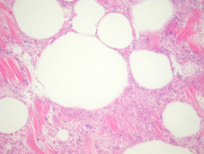
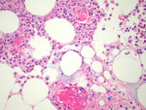
The expanded histological study shows marked reaction, accompanied by significant fibrosis and suppuration (Figure 3) along with extensive necrosis tissue necrosis (Figure 4). Even in hematoxylin-eosin stained sections, an overwhelming bacterial colonization Acute bacterial (Figure 4) is observed. Frequently, this process is accompanied by in the vessel wall. intravascular. intravascular thrombosis (Figure 5) and bacterial invasion of the vascular walls.
Key Pathological Findings in Necrotizing Fasciitis

Special Tests for the Diagnosis of Necrotizing Fasciitis
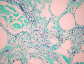
Gram stain is fundamental, as it confirms massive colonization by the organism causative organism (as illustrated in Figure 6). The case exemplified shows an abundance of Gram-positive cocci; however, it is crucial to remember that the morphology morphology and staining characteristics will vary significantly depending on the specific bacterial etiology.
Rigorous histopathological diagnosis and early identification of bacterial agents, especially in cases of necrotizing fasciitis, are essential pillars for instituting urgent and effective antibiotic treatment, which is decisive for patient survival.
A tissue biopsy or culture culture confirms the causal organism.
Intraoperative frozen section consultation is sometimes used for this disease. In addition to routine frozen section evaluation, touch preparations can be useful for identifying the presence of a microorganism. It is evident that an accurate diagnosis is fundamental for the appropriate surgical management of this very aggressive disease.
Differential Diagnosis of Necrotizing Fasciitis in Pathology
Panniculitis: Alpha-1-antitrypsin deficiency panniculitis and pancreatic panniculitis can manifest notable suppuration and necrosis, respectively. Organism identification and clinical correlation help establish the distinction.
Bacterial Overgrowth: Bacterial overgrowth is common in autopsy tissue autopsy and in unfixed tissue.
Juvenile NeutrophilicNeutrophilic Dermatosis: Tissue necrosis, thrombosis, and overwhelming bacterial overgrowth are generally not seen in these conditions.


