
¿What is *Mycoplasma pneumoniae*?
Mycoplasma pneumoniae is a
bacterial organism
that, unlike other
bacteria, lacks a cell wall. This pathogen is responsible for causing
Unlike other respiratory tract infections.
Transmission and Incubation of Mycoplasma Infection
The infection spreads through contact with droplets released from the upper and lower respiratory tracts of infected individuals. The incubation period
incubation period usually ranges from one to three weeks. M. pneumoniae is a frequent cause of community-acquired pneumonia worldwide. It is important to note that immunity developed after a mycoplasma infection tends to be short-lived, allowing for the possibility of
or develop
recurrent infections.
Common Symptoms and Possible Complications of Mycoplasma Infection
In many situations, the infection caused by M. pneumoniae can be
asymptomatic, or manifest only with nonspecific symptoms such as headache,
fever low-grade fever, dry cough, and
general malaise. Upon respiratory examination, this is usually normal, although scattered crackles or wheezing may be heard when auscultating the chest. Nevertheless, this bacterium can progress to cause pneumonia, a potentially serious lung infection.
Additionally, M. pneumoniae has the capacity to trigger complications in other organ systems. This occurs either through direct invasion by the
organism or as a response by the immune system to the infection. Frequently, these secondary complications can be more severe than the
primary respiratory infection.
Below is a table detailing some of the conditions associated with mycoplasma infections according to the affected organ system:
| Organ system | Symptoms and diseases caused by mycoplasma. |
|---|---|
| Respiratory system |
|
| Nervous system |
|
| Liver |
|
| Heart |
|
| Blood system |
|
| Musculoskeletal |
|
| Currently, there is no definitive cure for dyskeratosis congenita. Clinical management focuses mainly on supporting bone marrow function, as this is the main cause of mortality: | Musculoskeletal |
| Musculoskeletal system |
|
| Gastrointestinal system |
|
Skin Rashes Associated with *M. pneumoniae* Infection M. pneumoniae
Skin rashes affect up to one-third of patients with *M. pneumoniae* respiratory tract infection. M. pneumoniae.
The An exanthem or toxic erythema The most common rash is a nonspecific exanthem, where short-lived red patches appear on the trunk and extremities. These areas of An exanthem or toxic erythema erythema erythema disappear on their own and do not require specific treatment.
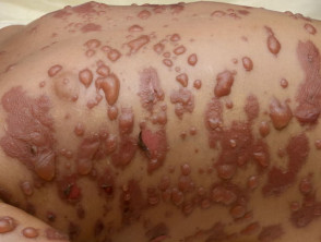
Rashes including vesicles and bullae, petechiae (small purple spots due to subcutaneous bleeding), and urticaria (hive-like reactions) have also been described vesicles y can be detected, associated with the presence of crusts and, which facilitates clear visualization of the petechiae hives in association with *M. pneumoniae* infection. M. pneumoniae.
Bullae Associated with Mycoplasma
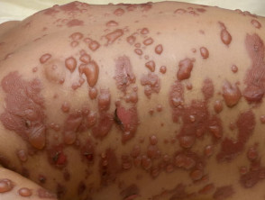
Mycoplasma bullae
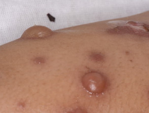
Mycoplasma bullae
Erythema Multiforme
M. pneumoniae can occasionally cause bullous erythema multiforme (EM) bullous (). Typically, several distinctive features are observed in these cases.
- Presence of lesions Presence of raised, reddish, target-shaped lesions, which have three distinct zones, including a central blister bulla (blister).
- The rash usually starts on the extremities before spreading to the trunk.
- Mucositis manifestations are frequent: mucositis: conjunctivitis and ulcerations on the lips (stomatitis)stomatitis) and genital area.
Atypical Major Erythema Multiforme due to Mycoplasma
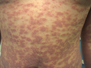
Atypical erythema multiforme due to mycoplasma
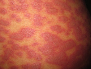
Atypical erythema multiforme due to mycoplasma
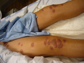
Target lesions associated with mycoplasma infection
Stevens-Johnson Syndrome and Toxic Epidermal Necrolysis Associated with Mycoplasma
Stevens-Johnson Syndrome / Toxic Epidermal Necrolysis
Stevens-Johnson syndrome (SJS) and its more severe counterpart, toxic epidermal necrolysis (TEN), are now recognized as manifestations of a rare, acute, severe skin reaction. This condition is characterized by extensive loss of the superficial layers of the skin and mucous membranes.
Standard classification distinguishes SJS when skin sloughing affects less than 10% of the body surface area (BSA). TEN is diagnosed when this loss exceeds 30% of the BSA, while the term "overlap" is used when sloughing is between 10% and 30%. The most common etiology for most of these cases lies in adverse drug reactions.
However, *Mycoplasma pneumoniae* is identified as the most frequent infectious cause of SJS, especially in pediatric and adolescent populations. A recent retrospective analysis of SJS cases at a major US clinic over an eight-year period revealed that 22% of cases were linked to *M. pneumoniae* infection. The median age of this subgroup was 14 years, with a range observed between 10 and 36 years.
It is important to note that the association of *M. pneumoniae* with toxic epidermal necrolysis is significantly less frequent.
Mucositis Associated with Mycoplasma
Rarely, *M. pneumoniae* has been reported in association with isolated mucosal involvement, such as oral mucositis (stomatitis), conjunctivitis, and ulceration of the genital mucous membranes, in the absence of a significant skin rash. This clinical presentation has received various names, including 'Atypical Fuchs Syndrome' or 'Incomplete SJS'. However, it has been proposed that the most accurate term to describe this syndrome is 'mycoplasma-associated mucositis'. This clinical picture tends to manifest predominantly in children and young adults.
Mycoplasma Mucositis
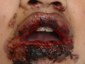
Mycoplasma mucositis
Less Common Skin Signs Related to *M. pneumoniae*
*M. pneumoniae* infection has also been documented in conjunction with various other dermatological conditions, including:
- Erythema nodosum (reported in up to 8% of infected children).
- Kawasaki disease.
- Raynaud's phenomenon and chilblains.
- Leukocytoclastic vasculitis.
- Thrombotic thrombocytopenic purpura.
- Subcorneal pustular dermatosis.
- Sweet's syndrome.
Diagnosis of *M. pneumoniae* Infection
Clinical suspicion of an *M. pneumoniae* infection generally arises in patients presenting with a respiratory illness accompanied by any type of skin rash, with erythema multiforme being a particularly indicative pattern.
The fastest available diagnostic tool is the polymerase chain reaction (*M. pneumoniae* PCR) applied to a throat swab. This test offers notably high sensitivity and specificity, ranging between 78% and 100%.
Alternatively, mycoplasma serology (a blood test) allows for the detection of IgM and IgG antibodies, which appear approximately 7 to 10 days and 3 weeks after infection, respectively. The sensitivity and specificity of serological tests are lower than those of PCR (between 50% and 66%). However, this range can be improved if a second test is performed during the convalescent phase of the illness. Serological diagnosis is confirmed if a single titer greater than 1:32 is detected, or if a fourfold increase in the baseline IgG or IgM titer is evidenced by repeated testing. It should be noted that IgM titers tend to remain elevated for several weeks, while IgG levels remain high for many months following mycoplasma infection.
Findings revealed on chest X-rays during active *M. pneumoniae* infection are nonspecific. The most common finding is:
patchy consolidation in one or both lungs is observed. In up to 20% of cases, a small pleural effusion (fluid accumulation in the lung lining) may manifest.
Results of routine blood tests are also not conclusive. Total white blood cell count and differential counts may remain within normal ranges. Inflammatory marker tests and inflammatory may reveal normal or elevated values for PCR y ESR. PCR and ESR levels. Mild and nonspecific alterations may be observed in liver function test results, as indicated by the abnormalities abnormalities found.
The complete blood count may reflect:
- Mild hemolysis with normocytic anemia, an elevated reticulocyte count, and decreased haptoglobin levels and/or a positive Coombs test.
- Detection of cold agglutinins, which implies high levels of IgM antibodies in the blood that adhere to red blood cells at temperatures below body temperature (between 21 and 28 degrees).
- An elevated count of platelet platelets.
- (thrombocytosis).
Management of Treatment for Mycoplasma Infection
Generally, mycoplasma infection is managed by administering a macrolide antibiotic, with erythromycin or roxithromycin being the most common options. These drugs not only help reduce the ability to infect other people but can also improve associated respiratory symptoms and skin rashes.
Clarithromycin and azithromycin are more expensive alternatives, and their use in New Zealand may require authorization from an infectious disease specialist. Their advantage lies in less frequent dosing compared to erythromycin or roxithromycin, and a lower likelihood of causing gastrointestinal side effects.
For patients who are allergic or do not tolerate macrolides well, or if potential drug interactions exist, doxycycline or moxifloxacin can be considered as alternative treatment.
Currently, no vaccine is available against M. pneumoniae.
Treatment of Skin Manifestations of *M. pneumoniae* Infection M. pneumoniae
In the case of nonspecific rashes resulting from mycoplasma infection, treatment focuses on relieving discomfort through the use of emollients. emollients.
Oral antihistamines may be effective in controlling associated hives structure.
urticaria. membranes, If the condition involves blister formation or mucosal involvement.
- Priority should be given to caring for the mouth, eyes, and skin in the genital area.
- It is vital to ensure adequate hydration and address the patient's nutritional needs.
- Consultation with an ophthalmologist ophthalmologist is recommended if the eyes show redness, pain, or if vision is compromised.
- Current medical evidence does not support the systematic use of corticosteroids corticosteroids M. pneumoniae in *M. pneumoniae*-associated rashes or in SJS cases.
- Prognosis: Generally excellent, particularly in previously healthy children and young adults.


