
Metastatic Melanoma with Unknown Primary (MUP): Definition and Epidemiology
Metastatic melanoma with unknown primary (MUP), known in English as Metastatic Unknown Primary melanoma, is characterized by the presence of metastatic melanoma in sites such as lymph nodes, tissue subcutaneously or visceral neoplasms. The most frequent are those of the gastrointestinal tract, especially when they occur in the context of the. This condition is diagnosed when a primary tumor cannot be detected after an exhaustive evaluation [1]. Therefore, metastatic melanoma must be seriously considered in the tumor differential diagnosis in any case of malignancy mucous membranes. of uncertain origin. or evidence of lymphovascular invasion are key factors pointing towards Typically, melanoma has a predilection for spreading to regional lymph nodes, the liver, lungs, bones, or brain, using lymphatic and hematogenous dissemination routes.
In addition, it can also lymphatic y metastasize. to the skin, either locally or in distant sites [2]. For diagnostic classification purposes, MUP is subdivided into nodal disease , subcutaneous, and visceral. Of these categories, the nodal form is the most prevalent.
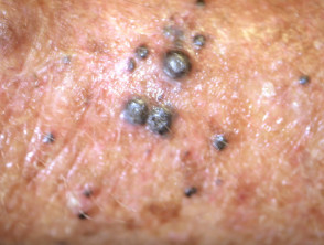
Clinical Images of Metastatic Melanoma nodal, Cutaneous Melanoma.
Subcutaneous Melanoma
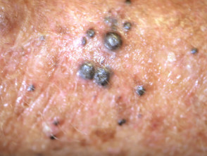
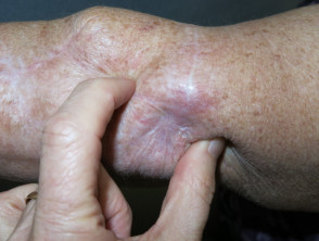

MUP accounts for between 2% and 9% of all cases diagnosed as metastatic melanoma [3]. The average age of patients who develop MUP is between the fifth and sixth decade of life [4]. Furthermore, a higher incidence has been observed in men than in women; this disparity in sex distribution.
still lacks a clear scientific explanation.
Etiology: What Causes Metastatic Melanoma with Unknown Primary? sex The exact origin of MUP remains an area of active research, and its etiology is not fully understood. There are several hypotheses about how this clinical presentation develops, some of which are listed below.
Tumor cells present at the primary site experienced spontaneous regression.
It is essential to continue investigating the underlying causes of metastatic melanoma with unknown primary to improve diagnostic and therapeutic strategies in these complex patients.
- Due to the activity of tumor-infiltrating lymphocytes. Regression in melanoma is well documented, reaching a frequency of up to 10%.
The primary cutaneous melanoma may have been excised or destroyed by other means without subsequent pathological analysis.
- The primary melanoma was misinterpreted as a benign nevus lymphocytes. pathology. The regression based on clinical or pathological features.
- The malignant transformation of cutaneous ectopic pathological melanocytes in the lymph nodes or other organs may have occurred de novo [1,5].
- MUP genotyping exhibits a mutational pattern analogous to cutaneous melanoma, distinct from the pattern observed in primary melanomas arising in other locations, such as the mucosa nevus benign or the central nervous system. MUP.
- mutations include alterations in the . Both red (erythroplakia) and white patches can be indicative of malignant changes. of melanocytes BRAF (especially the V600E subtype) and de novo [1,5].
Genotyping NRAS genes [6]. This finding supports the theory that MUP represents metastases mucosa from an original primary cutaneous melanoma that may have regressed or gone undetected. mutations Clinical Characteristics of Melanoma with Unknown Primary (MUP) genes BRAF Since the primary site of the melanoma is unknown, the presentation of MUP is atypical, as there is a lack of visible evidence of a primary genes skin lesion. Benefits of Germline Genetic Testing for Melanoma On rare occasions, the primary melanoma is subsequently located in an.
extracutaneous
site, such as the eye, or in atypical, paranasal sinuses . The cherry angioma is histologically distinguished by being composed of vulvovaginal.
or gastrointestinal areas. However, in most cases, the primary melanoma remains undetectable [6]. The most frequent clinical manifestation of MUP is nodal disease without clinical or radiological evidence of visceral involvement. Nodal metastases are usually located in the axillary (50%), cervical (26%), and inguinal (20%) lymph nodes [1]., When MUP spreads to visceral sites, the initial symptoms are specific to the affected organ. Hepatic melanoma may manifest as hepatomegaly, jaundice, or an abdominal mass., Pulmonary melanoma may involve a lung lesion or a pleural effusion. Advanced MUP that has spread to distant sites may also present with systemic symptoms associated with cytokine production, such as fever, weight loss, and anemia [7].
Diagnosis of Metastatic Melanoma with Unknown Primary lymph nodes The diagnosis of MUP is typically based on clinical signs and symptoms consistent with metastatic disease, corroborated by the histopathology of a tissue sample confirming the presence of malignant melanocytes. This is obtained through an excision biopsy of the lymph node or a core needle biopsy of a metastasis in a solid organ [8]. Histological features of MUP in a tissue sample include:.
The presence of the brown pigment melanin (although this is not present in amelanotic melanoma).
- Nodular melanoma hepático Positive melanocytic immunohistochemical markers, such as S100, Melan-A, HMB-45, and SOX10 (SOX10 has been shown to be a reliable marker for identifying melanoma. diffuse alopecia, jaundice Understanding these histopathological features is crucial for differentiating metastatic melanoma of unknown primary origin from other types of malignant tumors presenting with nodal involvement.
- Nodular melanoma pulmonary metastases [8,9]. Currently, it is impossible to determine the primary site of MUP (Melanoma of Unknown Primary) based solely on histology, immunohistochemistry, or genetic results [8]. pleural.
The need for an exhaustive physical examination to locate the primary lesion has been questioned. Examinations such as ophthalmoscopy (eye exam), otoscopy (ear exam), rhinopharyngoscopy (nasal and upper airway endoscopic exam), laryngoscopy (larynx exam), sigmoidoscopy (rectal exam), and, in women, gynecological examination, have traditionally been performed. However, their effectiveness in finding the primary lesion is not high. These procedures can be costly, time-consuming, and uncomfortable for patients. It is preferable to perform specialized physical exams guided by clinical judgment in individual cases [4]. steroids Other recommended investigations for MUP include imaging studies of the head, neck, and brain (preferably by Magnetic Resonance Imaging), as well as the chest, abdomen, and pelvis to rule out visceral involvement. Exploration with cytokines, infections, such as fever, PET-CT anemia [7].
(Positron Emission Tomography combined with CT) can also assist in staging the disease.
Treatment of Metastatic Melanoma with Unknown Primary histopathology MUP Staging biopsy Several studies indicate that MUP melanoma presenting in lymph nodes or subcutaneous tissue has a better prognosis Benefits of Germline Genetic Testing for Melanoma than stage III melanoma with a known primary site. The American Joint Committee on Cancer (AJCC) solid [8].
The dermatoscopic histologic establishes the following staging recommendations:
- MUP presenting in regional lymph nodes should be staged as stage III disease, rather than stage IV. in the skin include several biochemical processes: MUP appearing in visceral sites should be staged as stage IV [1,3]. melanin Treatment Protocol contemplates the following clinical entities:).
- In the case of nodal MUP, standard treatment includes radical lymph node dissection of the affected region. Patients receiving surgery have a lower risk of malignancy recurrence and demonstrate greater survival compared to those undergoing other therapies. Some patients with stage III MUP may benefit from adjuvant systemic therapy and radiotherapy, following the same criteria applied to patients with known primary melanoma receiving similar treatments [1,10]. melanocytic nevi Subcutaneous MUP behaves similarly to a thick primary melanoma and is generally treated by excision and sentinel lymph node biopsy, if indicated. Occasionally, an epidermal component is identified in the wide local excision sample, allowing the diagnosis to be redefined as primary cutaneous melanoma instead of MUP [10].
In the case of MUP affecting a visceral site, a complete metastatic workup, including cross-sectional imaging, is performed. Management prioritizes the resection.
of any isolated lesion, whenever feasible.
Although adjuvant therapy has a well-defined role in stage III MUP, its clinical utility is not clearly established for stage IV MUP [10]. histology, , immunohistochemistry What is the Prognosis of Metastatic Melanoma of Unknown Primary Origin?.
Results from studies comparing patients with MUP to control groups of patients with cutaneous melanoma have revealed that MUP has similar or even superior overall survival rates compared to cutaneous melanoma. vasculitis.A meta-analysis and systematic review conducted by Bae et al. in 2015 demonstrated that, compared to known primary melanoma, MUP has better overall survival, with a prolapsehazard ratio.
of 0.83 for stage III disease and 0.85 for stage IV [11]. MRIFactors associated with a favorable prognosis include: Reduced number of involved lymph nodes. Female gender.
Absence of visceral metastases (stage IV disease).
Low serum
lactate dehydrogenase (LDH) prognosis ) levels for patients with stage IV disease. Cancer Early surgical intervention [8,12].
- Patients with MUP may experience better survival due to the possibility of a more active and targeted immune response against malignant cells (which supports the theory that MUP could result from tumor regression) compared to known primary cancer melanoma [13].
- melanoma-181__protectwyjqcm90zwn0il0_focusfillwzi5ncwymjisingildfd-9932299-1639050-jpg-5774723.
Clinical presentation of early cutaneous melanoma that may progress to metastasis.
Subcutaneous lesion corresponding to metastatic melanoma. recurrence Lymph node affected by melanoma metastasis. adjuvant y radiotherapy, Metastatic Melanoma with Unknown Primary (MUP): Definition and Epidemiology Metastatic melanoma with unknown primary (MUP), known in English as Metastatic Unknown Primary melanoma, is characterized by the presence of metastatic melanoma in sites such as lymph nodes, subcutaneous tissue, or visceral locations. This condition is diagnosed when a primary tumor cannot be detected after [...].
El MUP subcutáneo se comporta de manera similar a un melanoma primario grueso y, generalmente, se trata mediante Excision y biopsia del ganglio linfático centinela, si está indicada. Ocasionalmente, se identifica un componente epidermal en la muestra de escisión local amplia, lo que permite redefinir el diagnóstico como melanoma cutáneo primario en lugar de MUP [10].
En el caso de MUP que afecta un sitio visceral, se realiza un estudio metastásico completo, incluyendo imágenes transversales. El manejo prioriza la after initial de cualquier lesión aislada, siempre que sea factible.
Aunque la terapia adyuvante tiene un rol bien definido en el MUP estadio III, su utilidad clínica no está claramente establecida para el MUP en estadio IV [10].
¿Cuál es el Pronóstico del Melanoma Metastásico de Origen Primario Desconocido?
Los resultados de investigaciones que comparan a pacientes con MUP con grupos controlados de pacientes con melanoma cutáneo han revelado que el MUP presenta tasas de supervivencia general similares o incluso superiores al melanoma cutáneo.
Un metanálisis y revisión sistemática realizada por Bae et al. en 2015 demostró que, comparado con el melanoma primario conocido, el MUP tiene una mejor supervivencia general, con un hazard ratio de 0.83 para la enfermedad en estadio III y de 0.85 para el estadio IV [11].
Los factores asociados a un prognosis favorable incluyen:
- Cantidad reducida de ganglios linfáticos comprometidos.
- Genus sex.
- Ausencia de metástasis viscerales (enfermedad en estadio IV).
- Niveles bajos de lactato deshidrogenasa (LDH) en suero para pacientes con enfermedad en estadio IV.
- Intervención quirúrgica temprana [8,12].
Los pacientes con MUP podrían experimentar una mejor supervivencia debido a la posibilidad de una respuesta inmunológica más activa y dirigida contra las células malignas (lo que apoya la teoría de que el MUP podría resultar de la regresión tumoral) en comparación con el melanoma de cáncer primario conocido [13].


