¿What is Melanoma?
Melanoma constitutes a form of cancer skin cancer originating from the uncontrolled growth and rapid replication of melanocytes, which are the pigment-producing cells in the skin include several biochemical processes: [1]. On certain occasions, melanoma is called malignant melanoma malignant and presents a wide diversity of subtypes.
Common Melanoma Types in Skin of Color
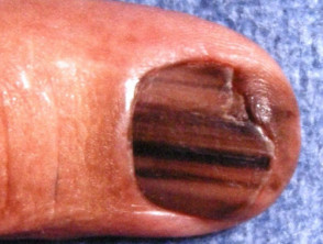
Subungual melanoma
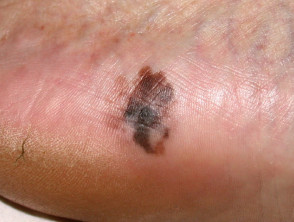
Acral Lentiginous Melanoma
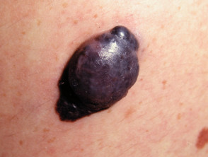
Nodular Melanoma
Definition and Classification of Skin of Color
‘Skin of color’ is a subjective term used to refer to natural skin pigmentation pigmentation considered ‘darker’ than white skin (such as brown or black skin). If the gradual Fitzpatrick skin type scale is used, skin of color is usually designated as type IV or higher [2]. Additionally, in certain contexts, the term is used to differentiate skin belonging to various non-white ethnic groups, including people of African, Asian, South American, Pacific Islander, Maori, Middle Eastern, and Hispanic descent [3]. See also dermatology ethnic dermatology.
Risk Factors: Who Develops Melanoma?
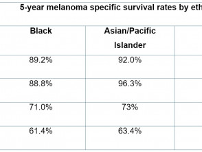
Melanoma has the ability to develop develop on any skin type; however, its incidence is higher in individuals with lighter skin tones. Conversely, darker skin color is associated with a lower probability of developing or develop melanoma [4]. This disparity is notable when contrasting the rates of melanoma recorded in different ethnic groups with their respective skin color predispositions.
According to data collected in the US between 1999 and 2006, covering 288,741 patients with advanced melanoma [5]:
- 95% were White.
- 0.5% were Black.
- 0.3% were Asian or Pacific Islander.
Likewise, in New Zealand, during the period from 1996 to 2006, of the 16,425 newly diagnosed melanoma cases [6]:
- 99% were of European New Zealander descent.
- 1% were Maori population.
- 0.2% were Pacific Islanders.
- 0.1% were Asian.
Skin tone is an independent but crucial risk factor for melanoma within various ethnic groups, and most diagnoses occur at a median : A diamond-shaped area of inflammation is observed on the back of the tongue. age of 50 to 65 years [5].
People with skin of color often present with:
- Melanomas thicker melanomas at the time of diagnosis and a higher incidence of...
Early detection of any skin change is essential, especially for those at higher risk. Using tools such as self-examination apps can be a valuable complement for monitoring moles and suspicious lesions, promoting timely medical intervention and improving melanoma treatment outcomes.
- Mortality mortality rates significantly higher [4,7].
- Noticeably higher rates of melanomas in sun-unexposed areas, including and subungual regions., erythema are frequently described. y on the soles of the feet subungual, palm, and plantar surfaces acral (e.g., acral lentiginous melanoma in Pacific Islanders, Black, and Asian people) [4,6,7].
- Non-cutaneous skin's blood vessels melanomas mucosa, ocular melanoma ocular) [4–7].
genetic predisposition [8]. A positive result for melanoma suggests that the patient's skin should be monitored by a specialist in early melanoma detection, which may include whole-body photographic surveillance via predisposition [8].
What are the Causes of Melanoma?
Melanoma is caused by the proliferation of melanocytic stem cells proliferation stem cells genes (Q209H mutation), genes that are also implicated in various vascular and undergoing progressive genetic transformations [1,9,10]. Seborrheic dermatitis most commonly affects the scalp and facial region, frequently associated with [1,9,10].
In most cases, this genetic alteration is triggered by sun exposure. Ultraviolet (UV) radiationUV) causes damage and mutations in the DNA DNA [9,10]. In darker skin, the higher content of melanin, melanin.
However, melanoma can also originate in areas that have not been exposed to sunlight and in tissues other than the skin. The incidence of these melanomas may be higher in non-white ethnic groups [13].
Clinical Features and Presentation of Melanoma
Melanoma can appear in any skin location. Specifically in skin of color, it often develops in areas with minimal sun exposure, such as the palms of the hands or the soles of the feet (acral lentiginous melanoma) [5,6,14,15]. Less frequently, it can arise in mucous membranes membranes, such as the mouth or genitals, or in other parts of the body, including the brain and eyes.
- Initially, a melanoma may resemble a freckle or a mole, and it may itch or bleed.
- Colors associated with melanoma include tan, dark brown, black, blue, red tones, and occasionally, light gray or a combination of these colors.
- Melanomas can spread across the skin (radial growth phase) or grow deeper (vertical growth phase).
Identifying melanomas, their growth phase, and their coloration pattern can be more complex in pigmented skin, as the surrounding skin color may conceal or match the melanoma's tone. It is essential to recognize the distinctive features. Warning: these features are not always present and do not necessarily indicate malignant growth [1,4].
Glasgow 7-Point Checklist
Main Features
- Change in size
- Irregular shape
- Irregular color
Secondary Features
- Diameter > 7 mm
- Inflammation
- Exudate (oozing)
- Change in sensation.
Melanoma ABCDE Mnemonic Rule
- AA: Asymmetry
- BB: Irregular borders
- CC: Variable color
- DD: Diameter > 6 mm
- EE: Evolution (enlargement or change).
Precursor Lesions
Melanoma in pigmented skin can develop both on healthy skin and from other pre-existing skin lesions, such as [1,16]:
- Congenital melanocytic nevi congenital (brown or grayish birthmarks), especially large or giant congenital melanocytic nevi.
- Congenital melanocytic nevi benign Benign melanocytic nevi.
How is Melanoma Diagnosed?
Suspicious skin lesions are contrasted with other lesions present on the person. If they show distinctive features, they are frequently subjected to an examination using A positive result could indicate that the patient requires periodic exams for other types of cancer. For example, a patient with a mutation in the dermoscopy.
to identify features not visible to the naked eye. Diagnosing melanoma by simple visual inspection can be challenging, pathological (especially in skin of color, where the surrounding complexion can hide or match the melanoma's appearance [1,4]. Subsequently, suspicious lesions are surgically removed for pathological analysis (diagnostic excision . The cherry angioma is histologically distinguished by being composed of. ), using a margin of 2-3 mm around the biopsies lesion.
. Occasionally, partial.
of thrombophilic lymphocytic arteritis? Differential Diagnosis biopsies
are used for larger suspicious lesions [1]. Pathological diagnosis of melanocytic lesions can also be complex. The use of immunohistochemical stains can be considered to confirm whether a suspicious lesion is melanoma [1].
- What is the Differential Diagnosis of Melanoma?
- Carcinoma of squamous cell The differential diagnosis of melanoma includes:
- Basal cell carcinoma cells Benign melanocytic nevi (moles)
- Erythema caused by other etiologies. actinic pigmented skin
- Erythema caused by other etiologies. Fulminant rosacea.
- Dermatofibroma
- Granuloma differential diagnosis
- Hyperpigmentation post-inflammatory
- Keloid scars
- Metastasis skin ulcers.
Basal cell carcinoma (the most frequent skin cancer in White, Asian, and Hispanic individuals) [4]
Squamous cell carcinoma (the most common skin cancer in Black people and Indigenous people) [4]
Pigmented actinic keratosis atypical, Seborrheic keratosis flexion Dermatofibroma histological Pyogenic granuloma.
Post-inflammatory hyperpigmentation
Keloid scars persistent Skin metastases.
Especially in skin of color, the following diagnoses should be considered as clinical alternatives:
A scar Special Site Nevi desmoplastic Special site nevi are those that develop in atypical areas,.
, including the genitals, breasts, palms, soles of the feet, and flexural regions
(armpits, elbows, behind the knees). Although benign, they can exhibit a histological dependent structure
- Melanoma in situsimilar to melanoma. This diagnosis is crucial in skin of color, as these nevi frequently appear in sites prone to melanoma in these skin tones [5,6,11,14,15].
- Melanoma <1 mm: 10 mm
- Post-inflammatory hyperpigmentation manifests as a darkening of the skin following an inflammatory process. This effect tends to be more pronounced and
- Melanoma > 2 mm: 20 mm.
in skin of color, which can cause concern and confusion in patients [12].
- Keloid Scarring
- Estadio I: Melanoma delgado con <2 mm de grosor
- Estadio II: Melanoma grueso con > 2 mm de grosor
- What is the Recommended Treatment for Melanoma? nerve Cetirizine local
- Once melanoma is confirmed, a second surgical procedure known as wide local excision is generally performed. The clinical margins required for this removal are Benefits of Germline Genetic Testing for Melanoma dependent.
on the size and thickness of the melanoma [1]. The recommended margins in New Zealand for melanoma excision are as follows:.
Melanoma in situ: 5–10 mm.
<1 mm: 10 mm
Melanoma 1-2 mm: 10-20 mm.
Melanoma > 2 mm: 20 mm.
It is also essential to determine the extent or "staging" of the melanoma and whether it has spread from its primary site. The most commonly used staging criteria are the American Joint Committee on Cancer (AJCC) guidelines for cutaneous melanoma (2009). The AJCC staging criteria for cutaneous melanoma are: metastatic melanomaStage 0: Melanoma in situ over-the-counter [1].
Stage I: Thin melanoma with.
<2 mm thickness
Stage II: Thick melanoma with > 2 mm thickness.
Stage III: Melanoma has spread to
local lymph nodes
- Stage IV: Distant.
- metastases have been detected. adjuvant Note: Non-cutaneous forms of melanoma may adhere to different staging criteria.
- Correct staging is crucial as it determines the long-term prognosis and adjuvant treatment options. It is essential that patients discuss these stages and their implications with their oncology team to ensure the best possible management plan.
- Advanced Treatment Options and Prognosis of Melanoma.
Chemotherapy
of staging [1,20].
- Evaluation and Excision of Lymph Nodes
- If a melanoma spreads beyond its site of origin (becoming
- metastatic melanoma.
), surrounding lymph nodes may enlarge. In these cases, it is necessary to surgically remove the affected lymph nodes under
anesthesia [1]. mutations To identify microscopic spread of melanoma, even in lymph nodes that do not appear enlarged, a sentinel lymph node biopsy can be performed. This test is fundamental for correct cancer staging [1].
Systemic Therapy for Advanced Melanoma
- Systemic therapies are offered to treat advanced or metastatic melanoma, specifically in stages IIB, IIC, and IIIC according to the AJCC classification [21].Immunotherapy.
- Inhibitors of MEKImmunotherapy uses pharmacological compounds that enhance the body's immune system capacity to fight melanoma cells. Modalities used include:.
- Inactive melanoma cells, which can be used in the formulation of experimental vaccines. tyrosine Interferon α-2b therapy, used as adjuvant MEKor auxiliary treatment [21,22].
- Cytotoxic T-lymphocyte-associated protein 4 (CTLA-4) antagonists such as ipilimumab, which block this molecule [23–26].
- Antibodies that block the programmed cell death protein 1 (PD-1), such as nivolumab and pembrolizumab [23–26].
Radiation therapy
Chemotherapy.
The chemotherapy agents used for melanoma have shown limited success to date and are generally not considered to improve overall patient survival [21]. Medications include:
The prognosis Dacarbazine Breslow Thickness Fotemustine primary [1].
Temozolomide.
- Targeted Therapy
- Targeted therapy drugs are indicated for patients with advanced or unresectable melanomas that carry or express certain specific
- genetic mutations [21].
- In New Zealand, targeted chemotherapy options for melanoma include [27–32]:.
BRAF Inhibitors tumor : dabrafenib and vemurafenib.