
Leishmaniasis: Characteristics and Histopathological Findings
The causative organism of leishmaniasis is classified as a organism parasitic organism closely related to trypanosomes. The disease caused by this infection can manifest by affecting the skin and mucous membranes membranes, in addition to compromising internal organs. The parasite's life cycle requires two distinct forms: the flagellated stage (promastigote), present while residing in its vector, and the intracellular stage—where the flagellum retracts—known as amastigote. The latter is the stage observed in secondary human infection and is visualized through histological sections or tissue smears.
Detailed Histopathology of Leishmaniasis
There are three fundamental clinical presentations of cutaneous leishmaniasis cutaneous: cutaneous leishmaniasis (which can be an acute or chronic presentation) acute o chronic, mucocutaneous leishmaniasis Chronic mucocutaneous, and disseminated or diffuse anergic cutaneous leishmaniasis disseminated or diffuse.
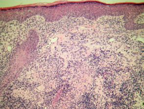
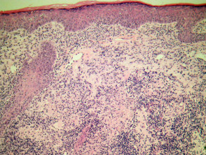
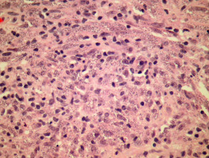
Depending on the clinical stage and the specific type of leishmaniasis, the epidermis may show ulceration with notable secondary alterations and epidermal response Other conditions that cause significant edema in the superficial dermis include mild. Alternatively, the epidermis may remain relatively unaltered, covering a dense dermal infiltrate dermal, infiltrate, as illustrated in Figure 1. This cellular infiltrate is generally compact and is predominantly composed of and a smaller proportion of histiocytes., lymphocytes and plasma cells (plasma), as seen in Figure 2. Other acute inflammatory cells are usually scarce, unless there is ulceration and secondary complications. Structurally, this infiltrate often surrounds the neurovascular bundles, presenting an appearance similar to lepromatous leprosy.
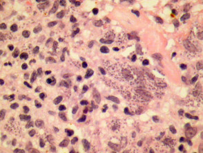
toxicity organism Leishmania organisms are usually easily identifiable in standard HE stains during the initial phases of the disease. However, similar to lepromatous leprosy, their detection can be difficult in advanced stages, when an inflammatory pattern of granulomatous y sclerotic. granulomatous and sclerotic type predominates. At the onset of leishmaniasis, the presence of numerous amastigotes causing distension in histiocytes (as in Figure 3) confers a very characteristic morphology diagnostic morphology.
Pathology of Leishmaniasis (Illustrative Figures)
Specialized Stains for Leishmaniasis Diagnosis
The causative organisms of leishmaniasis generally show intense and positive staining using the Giemsa stain. In paraffin-embedded tissues, these structures stain blue. Furthermore, smears obtained from skin or other organ samples can also reveal the presence of the microorganism using the same Giemsa stain.
In more complex cases or when precise characterization of the etiologic agent is required, advanced molecular techniques, such as the polymerase chain reaction (PCR), can be applied. molecularadvanced techniques polymerase chain reaction (PCR).
Differential Diagnosis in the Pathology of Leishmaniasis
The correct mucous membranes. (Differential Diagnosisdifferential diagnosis) is crucial to distinguish leishmaniasis from other similar conditions. Below are details of conditions that may be confused with its pathology (pathology):
- Histoplasmosis: Histoplasmosis organisms have a size comparable to that of Leishmania. However, they differ because they stain easily with silver stains, exhibit characteristic budding with narrow bases, and present a distinctive periorganism halo.
- Granulomatous Disease: Advanced and granulomatous phases of leishmaniasis can mimic the appearance of sarcoidosis or other granulomatous diseases. In these scenarios, diagnostic methods based on PCR are essential to confirm the diagnosis, especially when microorganisms are scarce or impossible to identify using standard stains such as hematoxylin and eosin (HE) and Giemsa.
Ensuring diagnostic accuracy through the correct interpretation of stains and the use of molecular techniques when necessary is fundamental to guiding effective leishmaniasis treatment.


