
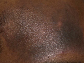
Key Differences in Caring for Colored Skin (Darker Skin)
The colored skin is generally defined as any skin type that is not white, with special attention paid to Fitzpatrick scale phototypes V and VI. This skin is distinguished by a higher concentration of epidermal melanin (the brown pigment), along with a more extensive distribution of melanosomes (the organelles containing melanin granules within melanocytes). This characteristic makes melanocytes and fibroblasts particularly reactive and hyperactive [Term: epidermal] [Term: melanin] [Term: pigment] [Term: melanosomes] [Term: granules] [Term: melanocytes] [Term: melanocyte] [Term: fibroblasts].
Common Manifestations in Colored Skin
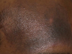
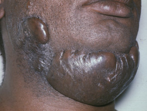
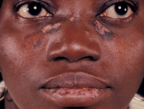
Biological Differences and Response to Damage in Colored Skin vs. White Skin
Because melanin is highly effective at absorbing and scattering energy from Ultraviolet Radiation (UVR), people with colored skin intrinsically experience less direct cellular damage after sun exposure and show fewer visible signs of photoaging compared to those with lighter phototypes [1,2].
However, this increased presence of melanin alters the inflammatory response. Colored skin almost universally develops pigmentary changes after suffering injuries or inflammatory processes (manifesting as post-inflammatory hyperpigmentation and the less common post-inflammatory hypopigmentation). In contrast, pigmentary changes are infrequent in white skin [3] [Term: pigmentary] [Term: inflammation] [Term: post-inflammatory] [Term: hyperpigmentation] [Term: hypopigmentation].
Melanin functions as a competitive chromophore (a colored molecule that absorbs transmitted energy), which increases the risk of adverse side effects after dermal procedures like laser therapy, as the main target absorbs a large portion of the energy [Term: chromophore] [Term: molecule] [Term: laser] [Term: UVR] [Term: inflammation] [4].
Additionally, people with colored skin have a greater predisposition to develop hypertrophic and keloid scars after an injury, compared to light-skinned individuals. This tendency is attributed to specific genetic factors that influence elevated fibroblast activity [Term: predominance] [Term: hypertrophic] [Term: genetic] [2,3].
Understanding these fundamental differences in biological response is essential for selecting effective and safe dermatological treatments. Successful management of conditions in colored skin requires an adapted approach that minimizes inflammation to prevent pigmentary alterations and the formation of keloid scars.
«`html
Indications and Considerations for Laser Treatment in More Pigmented Skin
Key Indications for Laser Therapy in Darker Skin
The fundamental medical applications of laser therapy remain consistent, regardless of skin tone. However, patients with colored skin frequently seek aesthetic intervention to treat hyperpigmentation and achieve skin tone uniformity, common problems in these phototypes [2].
Furthermore, laser technology is vital for the removal of thick, dark terminal hair. This procedure helps prevent dermatological conditions more prevalent in pigmented skin, such as pseudofolliculitis barbae, nuchal keloidal folliculitis, and folliculitis decalvans [5].
Safe Laser Hair Removal in High Phototypes
For hair removal in colored skin, the long-pulsed Nd: YAG (Neodymium, Yttrium, Aluminum Garnet) diode laser is generally recognized as the safest option.
- Its wavelength (1064 nm) is at the end of the spectrum where melanin absorption is lower.
- The Nd: YAG laser generates an adequate thermal injury in thick, dark hair while effectively protecting the epidermis [6].
- This device allows for a more gradual energy delivery, facilitating heat dissipation and surface cooling, which minimizes the risk of epidermal damage.
The Alexandrite laser (755 nm) has not been extensively evaluated in darker skin. Cases of blister formation have been documented in individuals with Fitzpatrick skin types V and VI [7]. Regarding the diode laser (800 nm), its application has shown to be mostly safe, with low complication rates reported, the most common being the appearance of transient blisters and transient and pigmentary alterations [8].
Acne Scar Treatment
While acne scars can be addressed with ablative lasers such as carbon dioxide or Er: YAG, their use is discouraged in phototypes V and VI. This is because the induction of thermal damage frequently causes post-inflammatory hyperpigmentation [9].
As an alternative, the non-ablative Nd: YAG laser presents a reduced side effect profile compared to ablative rejuvenation, offering comparable results with greater safety.
Tattoo Removal in Darker Skin
There is limited documentation on tattoo removal in pigmented skin. Successful removal of religious-themed black/blue carbon-based tattoos was reported in Ethiopian patients (skin types V and VI) using a Q-switched Nd: YAG laser Q-switched [10]. In that study, about half of the participants experienced mild post-inflammatory hyperpigmentation that resolved within a period of 2 to 4 months.
The difficulty in removing tattoos on darker skin lies in the presence of epidermal melanin. This melanin absorbs a large portion of the transmitted energy, preventing it from effectively reaching the ink deposited in the dermis [6].
Strategies to Optimize Laser Therapy Outcomes
Maximizing the success of laser procedures requires applying specialized techniques that prioritize the safety of pigmented skin.
Skin Cooling Methods
Cooling is essential to protect the epidermis from potential thermal burns during treatment [11].
- Contact cooling, which relies on conduction, can be active (using refrigerated sapphire or copper tips) or passive (such as ice application).
- Non-contact cooling employs cold air convection or cryocooling technologies.
Technical Treatment Protocols
It is crucial to rigorously monitor any sign of epidermal injury when working with colored skin.
- Generally, the Nd: YAG laser is preferred due to its longer wavelength and extended pulse durations, making it the most suitable device for these phototypes.
- It is advisable to perform initial test pulses or series to evaluate the immediate response of the laser on the patient's skin.
- Administering treatment in several short sessions usually proves more favorable for mitigating epidermal damage compared to single or overly prolonged procedures [12].
Patient Education and Expectations
It is essential that patients maintain realistic expectations about the results and are fully informed about the inherent risk of side effects and complications associated with any laser therapy [2].
```


