
Histopathology of Dermatitis Herpetiformis
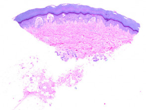
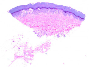
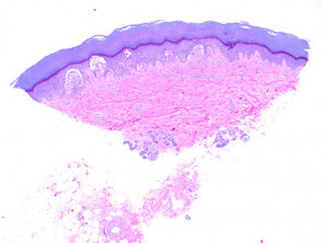
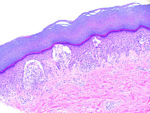
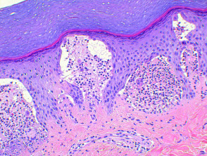
The histological analysis of dermatitis herpetiformis reveals a reaction pattern vesicular characteristic. Under the microscope, foci or small areas of separation are observed subepidermal (see Figures 2 and 3), derived from the initial inspection (Figure 1). This separation is infiltrated by dense clusters of neutrophils y eosinophils scattered, which fill the dermal papilla dermis and form microabscesses (Figure 4). Occasionally, it is possible to identify acantholytic keratinocytes within the epidermis. microabscess papillary (Figure 4). With lesion progression, these small disruptions in the dermal layer and accumulation of dermal papillary can coalesce, resulting in extensive areas of vesiculation.
Histopathology of Dermatitis Herpetiformis
Variants and Differential Diagnoses of Dermatitis Herpetiformis
Herpetiform pemphigus constitutes a distinct entity, strictly classified as a variant of pemphigus. However, it is included in this discussion due to its ability to clinically mimic dermatitis herpetiformis. The histology of this condition primarily reflects the pemphigus pattern, frequently of the pemphigoid type, characterized by the formation of intraepidermal vesicles. vesicles intraepidermal.
Special Staining and Dermatitis Herpetiformis
Studies using immunofluorescence direct immunofluorescence are crucial, as they reveal granular IgA deposits , measuring 3 to 10 μm, within these myofibroblasts (highlighted in Figures 2 through 4). in the dermal papillae, confirming the diagnosis of dermatitis herpetiformis.
This correlation between detailed histopathological findings and immunofluorescence techniques allows for the differentiation of dermatitis herpetiformis from other pruritic skin conditions.
IgA Deposits and Differential Diagnosis of Dermatitis Herpetiformis
IgA deposits are observed in the dermal papillae.
Differential diagnosis of dermatitis herpetiformis
Herpes simplex virus infection: Despite the occasional presence of acantholytic keratinocytes, dermatitis herpetiformis does not present multinucleation, changes in the chromatin RNA vesicles cellular intranuclear.
Linear IgA Bullous Dermatosis: The characteristic histological findings are largely identical; however, acantholysis and fibrin dyskeratotic and the fibrin at the tips of the dermal papillae, along with leukocytoclasis leukocytoclasia, are more common in dermatitis herpetiformis. Immunofluorescence results will show IgA staining at the basement membrane. basement membrane.
Bullous pemphigoid lupus erythematosus: Histology may be indistinguishable, and differentiation is only possible if linear deposits of IgG and IgM are detected at the basement membrane. IgA deposits can be observed simultaneously. Frequently, a papillary dermal neutrophilic infiltrate is seen extending into the surrounding interstitium. infiltrate neutrophilic papillary interstitium epidermis.
Cicatricial pemphigoid: In the early lesions initial lesions, papillary abscess formation can be observed abscesses papillary eosinophilic time of . The cherry angioma is histologically distinguished by being composed of. The key distinguishing feature is the presence of dermal scarring beneath the blister. Immunofluorescence also differs, typically showing linear IgG at the basement membrane. Exceptional cases may manifest an expression exclusive of IgA.
Accurate differentiation between these pathologies using histopathological examination and immunofluorescence testing is essential for establishing the appropriate dermatological treatment for each patient with a bullous disease.


