
Histology of Granuloma Annulare
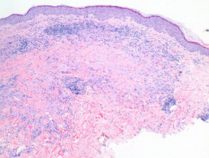
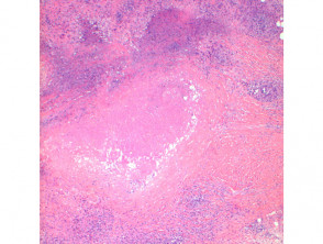
Histological analysis of granuloma annulare reveals a granulomatous inflammatory pattern located in the superficial and mid-dermis.
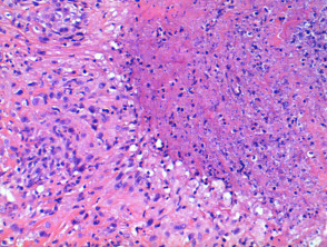
- Figure 1. A detailed inspection shows a palisade of histiocytes surrounding a focus of necrobiosis and an increase in mucin.
- Figure 2, Figure 3. The mid-dermis appears normal. It is common to observe multinucleated giant cells.
- Figure 4. In the surrounding dermis, a mild perivascular and interstitial lymphocytic infiltrate is appreciated, with scattered neutrophils and eosinophils. Plasma cells are scarce.
- Associated vascular fibrin deposition and nuclear dust, suggesting vasculitis, are infrequent findings.
Pathology of Granuloma Annulare
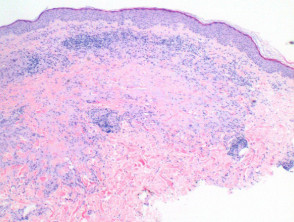
Figure 1
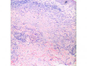
Figure 2
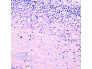
figure 3
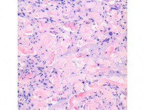
Figure 5
Histological Variants of Granuloma Annulare
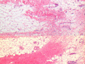
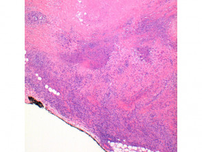
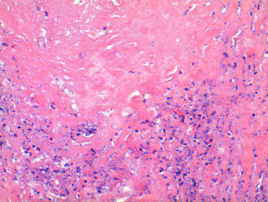
Subcutaneous Granuloma Annulare (SGA): In this variation, the inflammatory infiltrate is located predominantly in the deep dermis, extending into the subcutaneous tissue (Figure 5, 6). It is possible to observe large areas of necrobiosis accompanied by an increased count of eosinophils (Figures 7, 8, 9).
Pathology of Subcutaneous Granuloma Annulare
Special Stains and Syndromes Associated with Sebaceoma
There is a mitotically active variant; although mitotic activity is low, this variant can cause concern if it resembles a proliferative lesion proliferative. Caution is advised in these scenarios to rule out sarcoma epithelioid, as detailed below.
Perforating GA: In this case, elimination of the inflammatory infiltrate through the epidermis can be observed, epidermis, facilitated by a channel formed between the epidermal growths. changes.
Disseminated GA: Although the histology may be indistinguishable from typical GA, the infiltrate may be less dense and remain confined to the papillary dermis papillary dermis. In these situations, lichen nitidus is occasionally considered.
Interstitial GA: The low-power pattern is distinguished by showing an «occupied dermis,» lacking the characteristic necrobiotic and mucinous foci. Examination at higher magnification reveals an interstitial infiltrate of histiocytes that appear benign. Despite being less evident, an increase in mucin surrounding the inflammatory infiltrate is frequently appreciated. If there is significant presence of eosinophils and interface changes, a drug-induced granulomatous interstitial reaction should be considered.
Special Stains for Granuloma Annulare Diagnosis
To highlight the increase in mucins in the diseases, it is useful to employ mucin stains, such as colloidal iron and alcian blue.
Differential Diagnosis Key Differential Diagnosis of Granuloma Annulare
Necrobiosis lipoidica: It is distinguished by the presence of well-demarcated necrobiotic foci, but it lacks mucin deposits. An increased number of Plasma cells may be an important indication.
Nodule arthritisRheumatoid Nodule: The histological findings in this context are comparable to the subcutaneous variant of Granuloma Annulare. Key features for differentiating it from rheumatoid nodule include the observation of more extensive areas of eosinophilic necrobiosis eosinophilic and the absence of mucin deposits. Generally, the patient's clinical history is fundamental for distinction.
Epithelioid Sarcoma: It is crucial to reconsider this diagnosis when evaluating a Granuloma Annulare on the digits,. Although the low-power pattern can be simulated due to eosinophilic areas of necrosis, a detailed examination will always reveal an atypical atypical clear infiltrate. Immunohistochemistry Immunohistochemistry is definitive when demonstrating positivity for antigen Epithelial Epithelial Cytokeratin (CK) in lesions difficult lesions.


