
Extranodal NK/T Cell Lymphoma, Nasal Type: Characteristics and Pathology
The Extranodal NK/T cell lymphoma, nasal type, is a neoplasm driven by the Epstein-Barr virus (EBV). Usually, this condition primarily affects the upper respiratory tract (see also Lymphoproliferative Disorders Lymphoproliferative associated with EBV). Although skin involvement is usually secondary, in some cases the disease may manifest predominantly in the skin.
Histology of Extranodal NK/T Cell Lymphoma, Nasal Type
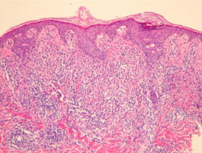
In Extranodal NK/T cell lymphoma, nasal type, histopathology typically reveals a dense dermal infiltrate infiltrate dermal, which frequently extends into the subcutaneous tissue subcutaneously (Figure 1). It is common to observe Inflammatory skin diseases examined through RCM can generally be classified into four main categories, allowing for more precise real-time diagnosis. of lymphocytes atypical changes within the epidermis. epidermis, atypical lymphocyte exocytosis within the epidermis, a feature that can closely mimic mycosis fungoides (Figure 2).
The dermis The dermis polymorphous, which may contain numerous histiocytes and eosinophils. and a smaller proportion of histiocytes. y eosinophils. The malignant cells vary in size, from small to medium, with some larger forms present (Figures 3 and 4). It is frequent for the infiltrate to involve the vascular walls (Figures 3 and 4), causing vascular vascular and nuclear debris pseudoinclusions (focally observable in Figure 4). Another recurrent pathological finding is the presence of necrosis. necrosis.
Images of the Pathology of Extranodal NK/T Cell Lymphoma, Nasal Type
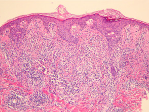
Figure 1
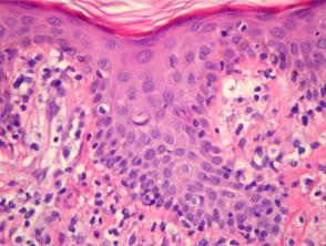
Figure 2
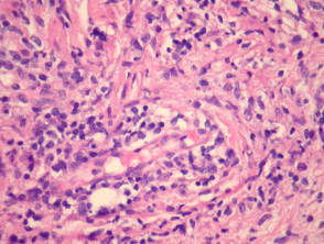
Figure 4
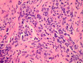
Figure 5
Understanding the histopathological features is essential for the differential diagnosis of atypical EBV-associated skin infiltrates. The polymorphous appearance and vascular involvement are key to distinguishing this lymphoma from other lymphoproliferative entities.
Specialized Analysis for Extranodal NK/T Cell Lymphoma, Nasal Type
Performing special stains is a crucial step in establishing the definitive diagnosis. At the lesion site, in situ hybridization with techniques for the Epstein-Barr virus (EBV) reveals positivity in the atypical cells. Generally, these cells show immunohistochemical positivity for markers such as CD56 and granzyme or TIA1. While some cases may express the T-cell marker, CD3, T-cell gene rearrangement studies usually yield negative results.
Differential Diagnostic Considerations for Extranodal NK/T Cell Lymphoma, Nasal Type
Other peripheral T-cell lymphomas and subcutaneous T-cell-like lymphomatous panniculitis can present morphological features resembling this type of lymphoma. If T-cell gene rearrangement studies are positive, along with positive T-cell immunohistochemistry, this would favor the diagnosis of a genuine T-cell lymphoma. The coexistence of positivity for EBV, CD56, and granzyme in a context of lymphoid malignancy is an infrequent finding in most other T-cell lymphomas, helping to delineate the diagnosis toward the NK/T subtype.


