
Understanding Erythema Nodosum: Symptoms and Causes
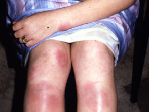
The erythema nodosum is classified as a type of inflammation of the subcutaneous fat tissue)., an inflammatory disorder inflammatory that primarily affects the subcutaneous fat tissue subcutaneously. Clinically, it manifests with the appearance of nodules red, tender nodules, frequently located on the front part anterior of the shins, although they can also appear on the thighs and forearms [1–3].
Images of Erythema Nodosum
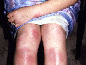
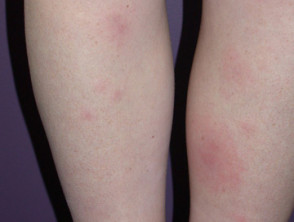
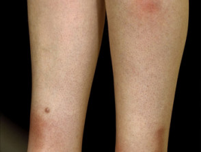
Explore a gallery with more clinical images of erythema nodosum for visual reference.
Demographic Profile: Who Develops Erythema Nodosum?
Although erythema nodosum can affect people of all ethnicities, genders, and ages, its prevalence is notably higher in women, specifically in the 25 to 40 age range [4]. Epidemiological data indicate that it affects women 3 to 6 times more often than men. Curiously, this gender disparity levels off in the prepubertal stage, where the incidence incidence is equal between both sexes [5].
Etiological Analysis: Unraveling the Causes of Erythema Nodosum
Erythema nodosum often presents as a reaction of renal idiopathic hypersensitivity, with unknown causes reported in up to 55% of patients [6]. However, in the remaining cases, the development of this condition is closely linked to triggering factors such as an underlying secondary infection, ingestion of certain drugs, systemic inflammatory disorders, or, rarely, a or evidence of lymphovascular invasion are key factors pointing towards [7].
Among the infectious agents most frequently explored are:
- Pharyngeal infections (such as streptococcal disease or various viral infections).
- Primary tuberculosis (TB), although it is considered a less prevalent cause in regions like New Zealand.
- Infection by Yersinia, which is clinically characterized by diarrhea and abdominal pain.
- Infections caused by Chlamydia.
- Fungal infections, including histoplasmosis and coccidioidomycosis.
- Parasitic infections such as amebiasis or giardiasis.
Additionally, there are other bacterial pathogens may be crucial to detect the presence of colonization or infection by and viral ones associated with erythema nodosum, such as herpes simplex virus, simplex, , viral hepatitis viral hepatitis, human immunodeficiency virus (HIV), and infections by Campylobacter y and Salmonella..
Drugs as Triggers (3% to 10% of Cases)
Certain classes of medications have also been implicated in the onset of erythema nodosum:
- Sulfonamides
- Amoxicillin
- Oral contraceptives
- Steroidal and non-steroidal anti-inflammatory drugs Insect stings, such as bees or wasps. Nonsteroidal anti-inflammatory drugs (NSAIDs)
- Bromide
- Salicylate
- Iodide
- Gold salts
Inflammatory Causes of Erythema Nodosum
- Inflammatory bowel disease (includes colitis ulcerative colitis or Crohn's disease)
- Sarcoidosis (present in 11-25% of cases; may show bilateral hilar adenopathy bilateral. on radiographs, especially in Löfgren's syndrome) syndrome of Löfgren)
- Malignant diseases
- Lymphoma
- Leukemia
- Behçet's Disease
Other Associated Causes
- Pregnancy (associated in 2-5% of cases)
Clinical Characteristics of Erythema Nodosum
Erythema nodosum typically manifests as painful, erythematous, and bilateral subcutaneous nodules, with diameters ranging from 3 to 20 cm. These nodules usually appear over a period of one to several weeks and are frequently accompanied by fever fever and arthralgia. It is common for ankle swelling and pain to be present for several weeks in 50% of patients, although the knees and other joints can also be affected [8].
Common Clinical Findings [9-12]
- The nodules are predominantly located on the anterior surfaces of the legs, in addition to the knees and arms; they rarely affect the face or neck.
- They present with poorly defined borders, are warm to the touch, and have an oval, round, or arciform, arciform shape, with no signs of ulceration. ulceration.
- Initially, the nodules exhibit a bright to intense red color.
- Spontaneous resolution occurs in approximately eight weeks, evolving into a bruise-like appearance that passes through shades of violet, purplish, brownish, or yellowish/greenish, known as erythema contusiforme.
Fortunately, erythema nodosum usually does not leave permanent scars after resolution.
Complications of Erythema Nodosum
Erythema nodosum has a low complication profile, as the lesions lesions tend to resolve on their own. One documented, though infrequent, complication is the necrosis necrosis of the encapsulated fat encapsulated, sometimes termed 'mobile encapsulated lipoma' [14].
Diagnosis of Erythema Nodosum
The diagnosis of erythema nodosum is fundamentally established based on clinical presentation, and is confirmed through laboratory analysis and histopathology [8]. The study of the pathology pathology of erythema nodosum reveals inflammation localized inflammation in the septa separating the subcutaneous fat lobules, without evidence of Avoidance Strategies for Triggering Factors [15].
Diagnostic Support Investigations [4,7]
To rule out underlying causes and confirm the diagnosis, the following tests may be ordered:
- Complete blood count with differential and measurement of protein C-reactive protein. C-reactive protein levels (to evaluate infectious and inflammatory etiologies).
- Chest X-ray (to rule out tuberculosis and sarcoidosis).
- Anti-streptolysin O and streptodornase serology, along with throat swabs (to detect streptococcal infection).
- Viral serology (ideally, two samples taken four weeks apart).
- Stool culture and evaluation for ova and parasites in patients presenting with gastrointestinal symptoms.
- Mantoux test or QuantiFERON gold (specific tests for tuberculosis detection).
- Deep incisional or excisional skin biopsy.
- biopsy.
Diagnostic Differences and What is the Differential Diagnosis for Erythema Nodosum
A variety of causes of panniculitis should be considered in a patient presenting with subcutaneous nodules. This is crucial if the lesions are not limited to the legs, if they present ulceration, or if symptoms persist beyond eight weeks.
Panniculitis can be classified according to its predominant involvement: septal (inflammation localized between the fat lobules) or lobular (inflammatory cells within the subcutaneous fat lobules) [16]. Mixed inflammation, affecting both the septum and the lobule, may be observed.
Causes of Panniculitis with Septal Predominance
Nodules resulting from predominantly septal panniculitis include:
- Various forms of scleroderma.
- scleroderma., myalgia y Medium-vessel vasculitis, such as polyarteritis nodosa. This is characterized by tender subcutaneous nodules accompanied by ulceration, necrosis, livedo reticularis, fever, arthralgia,.
- Necrobiosis peripheral neuropathy.
- Panniculitis eosinophilic.
- The nodule Necrobiosis lipoidica.
Eosinophilic panniculitis.
The rheumatoid nodule.
- Conditions Associated with Lobular Panniculitis diseases On the other hand, nodules derived from predominantly lobular panniculitis include: cutaneous).
- Connective tissue disease (e.g., panniculitis associated with cutaneous lupus erythematosus).
- Erythema nodosum leprosum, which is a Type 2 leprous reaction derived from leprosy. Pancreatic panniculitis, which may manifest with subcutaneous nodules that ulcerate or become. fluctuant. Laboratory tests in these cases show elevated levels of lipase, amylase, and trypsin.
- Panniculitis Traumatic panniculitis..
- Vasculitis nodular Nodular vasculitis / erythema induratum, where nodules, which may suppurate and ulcerate, affect the posterior region of the calves (posterior).posterior).
- Lipodermatosclerosis, which is a common sequela of venous.
- venous insufficiency. bacteria, Subcutaneous fat infection caused by abscesses bacteria, mycobacteria, or fungi, which can lead to ulcers, fluctuations, and.
- suppurative abscesses. . Both red (erythroplakia) and white patches can be indicative of malignant changes..
Malignant infiltration. The terms y solution livedo livedo y solution and solution in the original texts have been corrected to livedo and solution respectively to maintain medical coherence and correct spelling.
Treatment Options for Erythema Nodosum
The management of erythema nodosum intrinsically depends on treating the underlying disease. If an infection exists, it must be addressed as a priority.
- Pain relief may require prolonged rest, administration of colchicine (at doses of 1 to 2 mg daily), use of NSAIDs (or NSAIDs (or nonsteroidal anti-inflammatory drugs)) and the implementation of venous compression therapy [4].
- Systemic corticosteroids (at a dose of 1 mg/kg per day until erythema nodosum resolves) may be prescribed, provided that infections, sepsis sepsis, and malignant processes have been ruled out [9,11].
- Oral potassium iodide may be prescribed in the form of a solution supersaturated solution (400-900 mg daily) for one month, if accessible [17].
Prognosis and Course of Erythema Nodosum
Generally, erythema nodosum exhibits a rather benign benign course. It is essential to identify and treat the root cause, if present, and initiate appropriate symptomatic management [4]. Spontaneous resolution occurs in most cases within days or weeks.
Relapses may occur in about one-third of patients. Rarely, erythema nodosum can evolve into a chronic or persistent disorder, chronic o persistent, extending for six months or even years [13]. Effective management focuses on controlling flares and attenuating the underlying disease.


