
Understanding the Immune System and Autoimmunity
The immune system is composed of cells, molecules and structures designed to defend the body against external threats, protecting against Unlike other and monitoring any existing tissue damage [1].
The responses generated by this system are fundamentally classified into two categories: innate immunity innate and adaptive immunity.
Understanding Innate Immunity
Innate immunity refers to the generalized measures of generalized defense intended to protect the body against infection, including: secondary, including:
- Physical barriers, such as the skin and mucous membranes, membranes, which prevent the entry of most microorganisms [2]. microorganisms [2].
- White blood cells, exemplified by macrophages, macrophages, which have the ability to recognize microorganisms and secrete chemical substances to activate and attract other immune cells..
- toxicity Neutrophils, which surround and dissolve microbial invaders through a process known as phagocytosis, colloquially known as ‘cellular eating’ (cellular).
Generally, the reactions of the innate immune system manifest through inflammation [3].
Adaptive Immunity as Specific Defense
On the other hand, adaptive immunity involves immune responses that take longer to develop. These include the generation of specialized immune cells that produce undetectable designed to locate and eradicate a microorganism specific [1].
- In the cell-mediated immunity modality, T lymphocytes develop T lymphocytes that are trained to eliminate pathogens intracellular (such as viruses and bacteria) [4].
- In humoral immunity, the B lymphocytes, which are antibody producers, undetectable, fight pathogens found outside the cells (for example, bacteria encased in a polysaccharide capsule) [4]. polysaccharide) [4].
The main benefit of adaptive immunity is the creation of memory T lymphocytes and B lymphocytes (antibody producers). These cells have «recognized» a previous antigen and are prepared to respond specifically and rapidly if they encounter that virus or bacteria again in the future, eliminating it efficiently [3].
What Does Autoimmunity Involve?
Autoimmunity is defined as an immune response directed against the body's own tissues, typically involving T and B lymphocytes. The specific molecule or structure targeted by these immune cells is called an autoantigen [3].
This autoimmune response can lead to an autoimmune disease, autoimmune disease physiological function normal. However, autoimmune responses can also occur without causing pathological symptoms [3].
Antibodies that react against the antigens self are categorized as autoantibodies. autoantibodies. In certain autoimmune conditions, these autoantibodies are the direct trigger of tissue damage, while in others, they may be present without causing any injury [5].
Among the best-known autoimmune diseases are:
- Graves' disease (associated with hyperthyroidism).
- The arthritis Rheumatoid arthritis.
- Insulin-dependent nuclear Lesions of the Insulin.
- The systemic sclerosis Multiple Sclerosis.
- Systemic lupus erythematosus systemic [1].
Autoimmune Skin Diseases
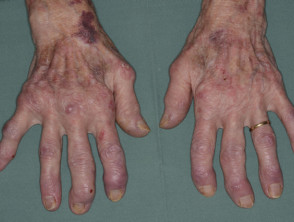
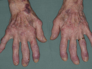
Systemic lupus erythematosus
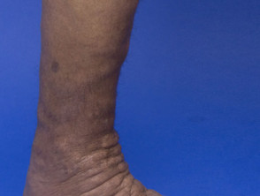
Pretibial myxedema in thyrotoxicosis
Among autoimmune skin diseases with blister formation, we find the following conditions:
- Pemphigus vulgaris and other variants of pemphigus.
- Bullous pemphigoid and other forms of pemphigoid.
- Epidermolysis bullosa acquisita.
- Dermatitis herpetiformis.
Manifestations of Autoimmune Bullous Diseases
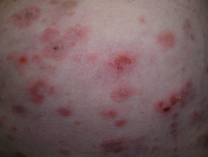
Pemphigus Foliaceus
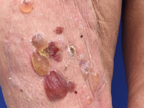
Bullous Pemphigoid
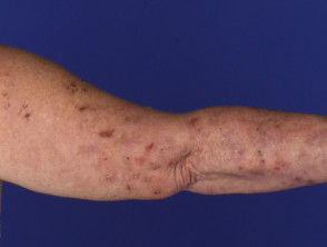
Dermatitis Herpetiformis
Additionally, autoimmunity and the subsequent dysregulation of the immune system are key factors in the development of several significant skin pathologies, including:
- Systemic sclerosis.
- Irritant contact dermatitis.
- Psoriasis.
- Vitiligo.
- Alopecia areata.
- Lichen sclerosus.
Details of Autoimmune Skin Diseases
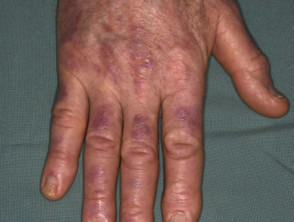
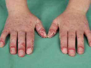
Gottron's papules in dermatomyositis
Localized vitiligo
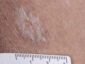
Extragenital lichen sclerosus
Understanding the various ways the immune system can mistakenly attack skin tissues is fundamental for accurate diagnosis and effective treatment of these complex autoimmune conditions.
Extragenital lichen sclerosus
Risk Factors and Impact of Autoimmune Diseases
Autoimmune diseases affect approximately 5% of the world's population [3]. It is crucial to understand who is most susceptible to these conditions.
Demographic profile of autoimmune diseases
- The vast majority of autoimmune diseases show a higher prevalence in women [3].
- There is an elevated risk of developing an autoimmune disease in individuals with a family history of these conditions [6].
- Certain geographic and socioeconomic patterns suggest a higher incidence; autoimmune diseases are more frequent in northern latitude populations and in higher socioeconomic groups [3].
- In contrast, autoimmune blistering skin diseases are rare and have incidence rates quite similar between men and women [7].
Mechanisms of Autoimmune Dysregulation
Autoimmunity manifests when the body's protective mechanisms, designed to prevent internal attacks, fail [5]. Several processes are key to maintaining immune tolerance:
- During the maturation of T lymphocytes in the thymus (a lymphoid organ key located between the lungs), T cells that react strongly against autoantigens are eliminated or inactivated [3].
- Circulating regulatory T cells play an essential role by actively suppressing other unwanted immune responses [5].
- B lymphocytes that show intense reactivity toward autoantigens may undergo receptor editing receptor, modifying their B cell receptors in an attempt to reduce autoimmunity [5].
- Both circulating T and B lymphocytes require co-stimulation provided by other immune cells to become fully activated [5].
Although the exact triggers of a specific autoimmune disease are often not fully determined, common risk factors include genetic aspects, disorders, previous infections, hormonal influences, and the use of certain drugs.
- Genetic factors disorders are often polygenic, implying that the combination of multiple genes increases the overall risk [1].
- Infections can initiate autoimmune processes, either by mimicking the structure of an autoantigen or by increasing the expression of co-stimulatory molecules [1].
- Some studies suggest that genes present on chromosome Y chromosome may offer male protection against autoimmune diseases, while estrogen estrogen susceptibility may influence the higher female susceptibility to suffering from them [1].
- Certain medications, including penicillamine, captopril, and vancomycin, have been associated with the ability to precipitating precipitate the onset of pemphigus vulgaris and pemphigus foliaceus [8].
Extensive clinical trials have consistently concluded that there is no evidence to support the claim that vaccines cause autoimmune diseases [5].
Diagnosis of Autoimmune Bullous Diseases
The autoimmune nature of this class of vesicular skin lesions is confirmed by positive direct tests. immunofluorescence Immunofluorescence microscopy reveals the presence hemosiderin of antibodies directly in the affected skin.
Pemphigus: Bullous Disorders Due to Cell Detachment
Pemphigus encompasses a group of rare blistering disorders, caused by circulating autoantibodies that target and firmly bind to adhesion molecules Labial present in the skin. This binding critically interferes with the ability of keratinocytes keratinocytes to remain cohesive.
causing intraepidermal intraepidermal blisters [9]. The main types of pemphigus include pemphigus vulgaris, pemphigus foliaceus, and paraneoplastic pemphigus [9]. paraneoplastic [9].
- Pemphigus vulgaris is distinguished by the appearance of blisters and and erosions erosions desmoglein 3, which is a crucial protein for keratinocyte cell-to-cell adhesion [10]. keratinocyte [10].
- Pemphigus foliaceus causes very superficial blisters, typically located on the trunk, scalp, and face. Identified autoantibodies target desmoglein-1, a molecule molecule responsible for holding dermal cells together [3].
- Paraneoplastic pemphigus manifests with blisters and ulceration ulceration or evidence of lymphovascular invasion are key factors pointing towards, malignancy proteins desmoplakin proteins [11].
Pemphigoid: Subepidermal Differences
The group of diseases known as pemphigoid includes bullous pemphigoid, mucous membrane pemphigoid and gestational pemphigoid. Unlike pemphigus, pemphigoid blisters are located in a subepidermal plane due to autoantibodies that specifically bind in the area of the edema epidermal-dermal junction [9]. epidermal-dermal [9].
- Bullous pemphigoid primarily affects the elderly, manifesting with large tense blisters filled with fluid and erosions. Frequently, these episodes are preceded by urticarial o eczematous plaques [12]. Antibodies against the bullous pemphigoid antigen (BP180), a hemidesmosome-associated protein involved in the adherence of keratinocytes to the hemidesmosome basal membrane basement membrane, and against bullous pemphigoid antigen 230 (BP230), present in basal keratinocytes [9]. cell [9].
- Mucous membrane pemphigoid is characterized by the recurrent appearance recurrent ulceration ulceration in the mucous membranes, particularly in the mouth and eyes, although it can also affect the skin [9]. Separation occurs deeper in the dermal-epidermal junction, which leads to scar formation. Various autoantibodies have been linked to different presentations of this condition, including antibodies against BP180, BP230, laminin 332, integrin alpha 6 and beta 4, and collagen type VII.
- Gestational pemphigoid affects women during pregnancy or in the period immediately following childbirth. Typically, it begins with intense itching, a hives hive-like rash rash centered around the navel, which can expand to practically the entire skin surface, excluding mucous membranes. Subsequently, it evolves into tense blisters similar to those observed in bullous pemphigoid [13]. Anti-BP180 antibodies may be detected. The primary site primary of autoimmunity is considered to be the placenta.
Other Uncommon Blistering Diseases
Dermatitis herpetiformis is an itchy, blistering skin condition that commonly affects the elbows, knees, and buttocks. It is associated with celiac disease and its symptoms significantly improve with the implementation of a gluten-free diet [14]. It is histopathologically characterized by blisters with a subepidermal deposit of immunoglobulin A (IgA) and a neutrophilic infiltrate. In this dermatitis, the antibodies of the autoimmune response target epidermal immunoglobulin A (IgA) and a infiltrate neutrophilic. neutrophilic infiltrate When acute hives is caused by reactions similar to serum sickness (such as those following blood transfusions or certain medications), it may be accompanied by ecchymosis (bruising), fever, transglutaminase, involved in clotting tissue coagulation [15].
Linear IgA bullous dermatosis dermatosis is an extremely rare autoimmune blistering disorder, susceptible to being acquired or drug-induced (such as vancomycin). Blisters may occasionally be arranged in annular configurations (known as the 'string of pearls'......
... signature) [9]. There is a subepidermal deposition of IgA antibodies that targets a fraction of the BP180 antigen, type VII collagen, or other essential basement membrane proteins.
Epidermolysis bullosa acquisita is equally rare. In its classic presentation, blisters and erosions originate in areas of minor trauma [9]. In the case of epidermolysis bullosa acquisita, the autoimmune response specifically attacks type VII collagen, located in the basement membrane zone of the skin and mucosa. mucosa.
Bullous systemic lupus erythematosus represents a rare presentation, characterized by subepidermal blisters in patients already suffering from systemic lupus erythematosus.
Diagnosis of Autoimmune Dermatological Diseases
a biopsy biopsy is generally required to obtain a definitive diagnosis of any autoimmune skin disease, although characteristic clinical morphology can strongly suggest a specific condition [9].
Blood tests that may be useful in the diagnostic process include:
- Circulating cutaneous autoantibody tests via indirect immunofluorescence [14]
- Specific tests for celiac disease, such as the detection of tissue transglutaminase IgA antibodies
- Determination of non-specific and inflammatory inflammatory markers C-reactive Protein measurement))
- A broad autoimmune screen (e.g., detection of pseudoinclusions antinuclear antibodies).
bacterial infection Acute bacterial or infections caused by the herpes virus.
Differential Diagnosis of Autoimmune Bullous Skin Rashes
Skin rashes with blister formation associated with systemic diseases often manifest with fever fever
- The syndrome Stevens-Johnson syndrome / toxic epidermal necrolysis toxic
- Staphylococcal scalded skin syndrome
- Varicella-zoster infection
- Herpetic infection disseminated, eczema herpeticum (eczema herpeticum) or herpes simplex in simplex immunocompromised patients immunocompromised
- Bullous variant of systemic lupus erythematosus
- Acute febrile neutrophilic dermatosis acute y febrile.
Other generalized conditions that cause blisters and share symptomatology with autoimmune skin diseases are:
- Bullous impetigo
- Miliaria crystallina [14].
The appearance of blisters on mucous membranes is also a key symptom symptom in the following conditions:
- Erythema multiforme [90]
- Cutaneous viral infections (e.g., Herpes simplex, herpes zoster or stomatitis enteroviral stomatitis)
- Livedo Aphthous ulcers
- Behçet's syndrome [14].
Treatment of Autoimmune Skin Conditions
The therapeutic approach for autoimmune skin diseases varies significantly depending on the specific pathology diagnosed.
The fundamental treatment for pemphigus and pemphigoid is based on the use of corticosteroids corticosteroids and immunosuppressive therapies [16]. [16].
On the other hand, dermatitis herpetiformis is successfully managed by administering dapsone supplemented with a strict gluten-free diet [15].
Determining the appropriate treatment protocol is crucial to control symptoms and prevent long-term complications in these rare dermatological conditions.


