
Apocrine Mixed Tumor: Characteristics, Histology, and Differential Diagnosis
The apocrine mixed tumor, also known as apocrine chondroid syringoma, typically manifests as slow-growing dermal nodules, whose appearance can be nonspecific. These skin lesions are most frequently located on the skin of the head and neck.
Detailed Histology of the Apocrine Mixed Tumor
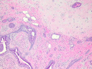
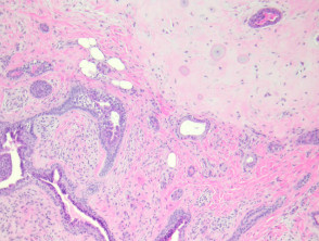
Apocrine mixed tumors present macroscopically as a well-circumscribed dermal mass. These tumors are characterized by their dual structure, composed of a mesenchymal component, frequently chondroid in nature, and an epithelial component (see Figure 1).
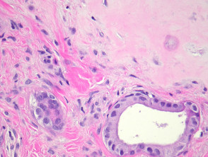
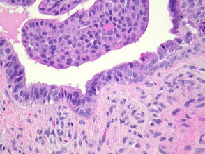
The cartilaginous portion is mature, showing well-differentiated chondrocytes and a grayish or silvery matrix (Figure 2). For its part, the epithelial tissue is bland and is organized predominantly into two cell layers: a lining luminal layer and an adjacent myoepithelial layer (Figure 3).
Pathology Images of Apocrine Mixed Tumor
Complementary Studies for Apocrine Mixed Tumor
Generally, performing additional special studies is not required to establish the diagnosis of a benign apocrine mixed tumor.
Differential Diagnosis of Apocrine Mixed Tumor
The differential diagnosis is crucial, especially when considering the possibility of malignant transformation:
- Malignant Apocrine Mixed Carcinoma: The presence of marked cellular atypia, numerous mitoses, and invasion of lymphovascular spaces suggests malignant transformation, although this is an unusual event. Given the rarity of reported metastases even in bland-appearing variants, some experts recommend complete surgical excision of all these tumors.
- Ecrine Mixed Tumor: This tumor is distinguished by glandular spaces of relatively regular size, lined by thin epithelium and surrounded by a fibromucinous stroma.
- Pleomorphic Adenoma: Salivary gland tumors, specifically pleomorphic adenoma, can be morphologically identical to apocrine mixed tumors. The location in the parotid gland or minor salivary glands generally facilitates the distinction between these entities.
A correct histopathological evaluation is essential to differentiate benign apocrine mixed tumor from its malignant counterparts or similar adnexal gland lesions.


