
Key Concepts in Digital Microscopy: Whole Slide Images
Whole Slide Imaging (WSI) involves the software-based manipulation of digitally scanned tissue sections under various magnification levels. This essential technique essentially allows the observer to perform a detailed zoom into specific areas of interest, emulating the experience of examining glass slides under a traditional microscope, reaching magnifications up to ×40. microscope traditional, reaching magnifications up to ×40.
Whole slide images are also known by the term microscopy virtual.
Visualization of Whole Slide Images
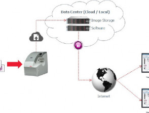
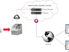
Courtesy of Colin Doolan, Leica Biosystems
Advantages of Implementing Whole Slide Microscopy
There are multiple benefits to opting for digital whole slide images for microscopic examination microscopic of specimens pathology, highlighting the following aspects:
- Ease of file duplication and distribution.
- Greater durability: No risk of physical deterioration or breakage.
- Optimized management: Relatively simple storage, cataloging, and administration.
- Remote accessibility: Available via the internet, eliminating geographical barriers.
An important milestone was when the U.S. Food and Drug Administration (FDA) approved the commercialization of these slide scanning systems for use in pathological diagnosis pathological in April 2017.
The Dermatopathology benefits enormously from the implementation of WSI for diagnosis, given the high concurrence of cases, the manageable volume of slides, and the small size of samples in this specific discipline.
Educational and Diagnostic Applications of Virtual Microscopy
Training, Assessment, and E-Learning
Currently, educational institutions have computer equipment that is more affordable than microscopes physical, facilitating the transition to digital.
- WSI offers students and residents access to a vast library of rare case slides, which would otherwise not be available, while also protecting valuable glass slides from wear or breakage.
- It ensures that all participants analyze exactly the same slide, ensuring uniform exposure to critical information.
- Advanced software allows for complete image rotation (360 degrees), facilitating review from any perspective, crucial for evaluating the orientation of the epidermis in dermatopathology slides, a significant limitation in traditional teaching.
- Software tools allow for the integration of annotations (circles, arrows), clinical photographs, results of A positive result could indicate that the patient requires periodic exams for other types of cancer. For example, a patient with a mutation in the or radiological images, making this platform an ideal resource for teaching the necessary clinicopathological correlation. By adding detailed discussions, multimedia content, external links, and bibliographic references, the quality of teaching is substantially elevated.
- The utility of images as fundamental tools for teaching and training.
- Whole slide images optimize autonomous learning (e-learning) and, by integrating additional software, facilitate immediate real-time feedback. This allows participants to continuously self-assess their performance. Once digitized, auxiliary material and images are available 24/7 for later review and study.
- The digitization of whole slides facilitates pathology assessments without the need for physical microscopes. In fact, by 2020, the American Board of Dermatology Dermatology exclusively adopted whole slide images for its certification exams, completely eliminating the use of microscopes and glass slides.
Continuing Medical Education (CME) and External Quality Assurance
Whole slide images are also valuable in contexts of continuing medical education (CME) or continuing professional development (CPD), as well as in external quality assurance (EQA). Integrating assessments with embedded multiple-choice questions facilitates obtaining CME credits online. Accumulating CME credits is increasingly crucial for specialists: in some nations, such as currently in Europe, this can translate into salary increases; in others, it is a mandatory requirement for license renewal and maintenance of specialty certification, as already occurs in the U.S. along with mandatory certification exams.
Globally, digital pathology has proven to be a robust platform for CME, CPD, and EQA programs. Several key organizations, including the College of American Pathologists, the American Society for Clinical Pathology, and the Royal Australasian College of Pathologists, actively employ digital pathology to conduct accredited CME exercises. Between 2013 and 2015, the European Academy of Dermatology and Venereology provided its members with a quarterly clinicopathological self-assessment module using whole slide images. For its part, the American Society for Dermatopathology presents a monthly interactive case study that also incorporates these images. Furthermore, several EQA programs for pathology subspecialties in the United Kingdom utilize digital pathology. The Nordic Institute of Virtual Dermatopathology organizes annual Nordic courses, online modules, and CME exams using whole slide images and associated software.
Effective Consultation and Collaboration
Consultation between pathologists and clinicians worldwide is greatly facilitated. Appropriate software allows for the incorporation of the following data along with slide information:
- Gross morphology gross
- Relevant clinical photographs
- Dermoscopic images dermoscopic,
- Dermoscopic images radiological images
- Specific annotations
- Insertion of comments or notes.
Dermatopathology Sample Image *
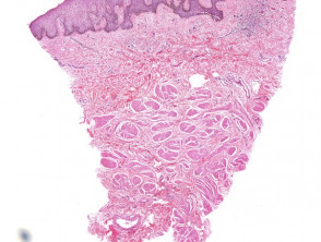
Sample image of a whole slide image
* Click here to view the original slide in the digital slide viewer
Storage of Scanned Images for Archiving, Tumor Boards, and Research
Several institutions are adopting whole slide images for archiving and document management purposes.
- Given the growing scarcity of physical space to store histopathology slides carcinoma, scanning relevant samples and subsequent disposal of the original glass slides (retaining tissue blocks) has become an established viable alternative. This practice also significantly reduces the operational costs associated with staff responsible for retrieving physical slides.
- Mass storage of collections or software, including digital slides, can...
The implementation of digital pathology through whole slide scanning not only optimizes archive space and reduces operational costs but also enhances collaboration and academic rigor through continuing education and global consultation. These digital resources are becoming the essential standard for certification and modern practice in dermatopathology.
Benefits and Applications of Whole Slide Imaging
Ease of Implementation and Storage
- Implementation is facilitated in data centers (whether cloud-based or local), while individual collections can be easily safeguarded or backed up on hard drives.
- Subsequently, digital slides are easily retrieved for use in research or tumor case discussions.
Integration into Electronic Medical Records (EMR)
- Digital pathology slides can be attached directly to EMRs, optimizing consultation and streamlining information transfer processes.
- In the future, the ability to access these files remotely via specialized applications is anticipated.
Pattern Recognition Algorithms as a Diagnostic Tool
- Whole slide images enable quantitative analysis, both manual and computer-assisted, of the diagnosis and prognosis of protein and genetic prognosis of biomarkers protein y disorders.
- Various algorithms already have FDA (Food and Drug Administration) approval for clinical use, and more solutions are continuously being developed.
Challenges of Whole Slide Imaging
The potential disadvantages associated with adopting whole slide imaging may include several technical and economic factors:
- The availability of histopathology scanners histopathology is not universal in all centers, although their presence is increasing. Alternatively, it is feasible to send slides to commercial centers for scanning via a one-time payment.
- A high-speed Internet connection is indispensably required to view large files (the previous example required 512 Kbps); this connection speed is already standard in developed nations.
- There may be incompatibility issues between digital slides scanned by different scanner manufacturers and the software used for their viewing. However, current software is designed to be compatible with most digital image formats, regardless of their origin.
- Some viewing software that relies on FlashPlayer™ is not compatible with some tablet-type devices, such as the iPad™.
- Specialized software for whole slide imaging is expensive, as are associated hosting and managed services. The expected lifespan of such software might be limited.
Research on the Reliability of Whole Slide Imaging
Exhaustive diagnostic accuracy studies have been conducted using whole slide images. These studies have contrasted the use of traditional glass slides versus digital ones, and have also compared conventional photomicrographs with digital versions. Some relevant references are listed below; this is not an exhaustive list of available literature.


