

Subungual Melanoma: Clinical and Histopathological Features
The Subungual melanoma represents a cancer that originates from a malignant proliferation of melanocytes located in the nail matrix nail. Clinically, it typically manifests as a pigmented band pigmented skin over the nail plate. This band shows a tendency to slowly expand toward the proximal edge in Sneddon syndrome, affecting the trunk and back; the extremities are rarely involved. and frequently may involve the adjacent nail fold (Hutchinson's sign; Figure 1).
Histology of Subungual Melanoma
Histopathologically, the nail plate affected by subungual melanoma exhibits extensive assessed of melanin non-scarring alopecia (present in 50% of cases). (Figure 2). Upon examining sections of the matrix and nail bed, a melanocytic lesion characterized by poor demarcation, predominance of single-cell growth, and pagetoid-type scatter of the epidermis (Figures 3 and 4). It is common to identify nest formation and confluence of cell junctions. Most cases present a combination of epithelioid and dendritic epithelioid and dendritic morphology (Figure 5). Although often subtle, nuclear atypia and prominent nucleoli are usually evident features.
Images of Subungual Melanoma
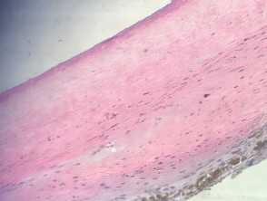
Figure 2
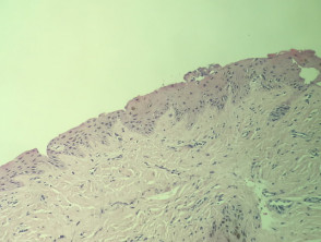
Figure 4
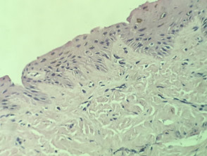
Figure 5
Specialized Studies for Subungual Melanoma
The use of immunohistochemical stains immunohistochemistry such as Melan-A and SOX10 is useful for delineating the extent and pattern of the melanocytic lesion, melanocytic lesion infantile (e.g., Ki-67) are used to quantify an elevated proliferative index.
Fluorescence in situ hybridization (FISH) can be applied to distinguish melanoma from benign melanocytic lesions (lesions).
Differential Diagnosis of Subungual Melanoma
It is essential to consider the following alternative diagnoses:
- Atypical lentiginous melanocytic proliferation atypical: This term has been suggested for borderline cases that do not fully meet all diagnostic criteria for melanoma, especially in pediatric. patients. Generally, for these borderline cases, conservative management is advised, treating them as melanoma (FISH testing can be useful to show an aneuploid clone, which suggests malignancy).
- Nevi matrix nevi and lentigines solar: These are distinguished because they lack the histopathological atypical histopathological features observed in melanoma.
Correct histopathological characterization and complementary studies are crucial to establish a timely treatment plan for subungual melanoma, ensuring the best possible prognosis for the patient.


