
Subcorneal Pustular Dermatosis (Sneddon-Wilkinson Disease): Key Symptoms
The subcorneal pustular dermatosis, also known by its historical name as Sneddon-Wilkinson disease, is an uncommon dermatological condition. Its main characteristic is the sustained appearance of pustules that can persist for prolonged periods, spanning months or even years.
It is important to note that, on certain occasions, the clinical presentation might lead to a subsequent reclassification as Generalized Pustular Psoriasis; however, the latter condition tends to evolve with more severe systemic and cutaneous symptoms.
Characteristics and Clinical Manifestations of the Disease
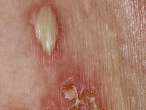
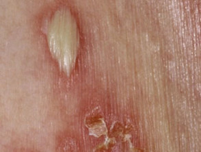
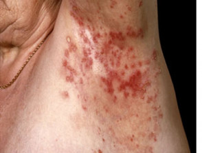
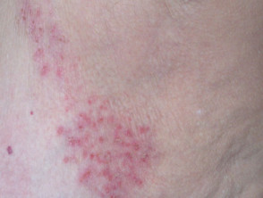
Subcorneal pustular dermatosis is clearly identified by the presence of multiple soft pustules that arise on the skin surface. These lesions are usually located predominantly on the trunk, particularly in fold areas such as the armpits and inguinal regions. Although they can emerge on apparently normal skin, they are frequently observed within an patch of erythema, meaning an area of skin redness.
The evolutionary cycle of these pustules is inherently self-limiting: they resolve spontaneously in a few days, leaving behind a layer of scaling, before a new outbreak occurs, characterized by desquamation, relapse and the formation of new pustules. Despite this lesional activity, these lesions generally only cause mild itching, with the patient remaining asymptomatic systemically. The clinical pattern is recurrently cyclical: outbreaks can reappear for weeks, disappear for months or even years, and then return. This cyclical nature of recurrence can persist for a long time.
From an epidemiological perspective, subcorneal pustular dermatosis disproportionately affects women more than men, with the typical age group for onset being individuals over forty years old.
Visual Gallery: Subcorneal Pustulosis and Sneddon-Wilkinson
Subcorneal Pustular Dermatosis
Subcorneal Pustular Dermatosis
Subcorneal Pustular Dermatosis
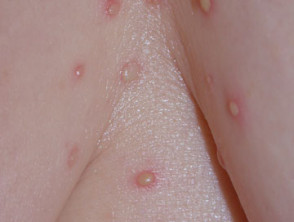
Subcorneal Pustular Dermatosis
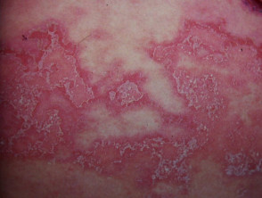
Subcorneal Pustular Dermatosis
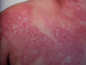
Subcorneal Pustular Dermatosis
Understanding the cyclical course and typically truncal distribution of these lesions is crucial for the correct diagnosis of Subcorneal Pustular Dermatosis and to differentiate it from other more aggressive pyodermas. If you or someone you know presents with these persistent skin characteristics, it is essential to consult a dermatologist for an accurate evaluation.
«`html
Etiology and Diagnosis of Subcorneal Pustular Dermatosis
Etiology: Unknown Causes and Associated Conditions
To date, the specific cause that originates subcorneal pustular dermatosis remains unknown.
However, an association with certain underlying medical conditions has been documented. The most frequently reported include:
- The IgA monoclonal gammopathy (abnormalgammopathy due to immunoglobulin accumulation).
- Myeloma with paraproteins (dysproteinemia) in the blood.
- Multiple myeloma.
- Pyoderma gangrenosum.
Other less frequently associated conditions include rheumatoid arthritis, lupus erythematosus, hyperthyroidism and hypothyroidism, polycythemia vera, and SAPHO syndrome (which involves synovitis, acne, pustulosis, and osteitis).
The presentation of subcorneal pustular dermatosis can be very challenging to differentiate from Generalized Pustular Psoriasis and Acute Generalized Exanthematous Pustulosis. Both disorders appear to be closely linked, making a correct differential diagnosis essential.
Diagnostic Evaluation and Procedures
A skin biopsy is frequently performed to confirm the diagnosis of the condition. This procedure is fundamental for histopathological confirmation.
Requested blood tests usually include a general examination, such as a complete blood count, calcium measurements, and liver function tests, in addition to protein electrophoresis to detect any gammopathy present in the patient.
Treatment Options for Subcorneal Pustular Dermatosis
The main objective of treatment is the prevention of complications associated with the condition. Dapsone is generally successful, achieving resolution of the lesions in approximately one month. In some cases, continuous maintenance therapy with a reduced dose may be necessary to control the disease long-term.
Other therapeutic alternatives are available if dapsone is not effective or tolerable, such as:
- Acitretin.
- Sulfapyridine or sulfamethoxypyridazine (the latter is not available in New Zealand).
- Phototherapy, including UVB and PUVA treatments.
- Colchicine.
- Cyclosporine or other immunosuppressive agents such as mycophenolate mofetil.
- Biologic therapies targeting inflammatory mediators, including infliximab and adalimumab.
Various agents have been successfully used in individual patients. It is important to note that systemic steroids are usually ineffective and, in fact, have the potential to precipitate or cause an outbreak of subcorneal pustular dermatosis, so they should be used with caution.
It is crucial that patients undergo long-term follow-up, including periodic evaluations of serum protein electrophoresis and immunofixation. This is because paraproteinemia or myeloma can develop years after the initial presentation of the dermatosis.
```


