
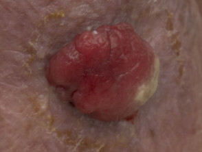
![of the primary lesion, ensuring clear margins [1].](https://dermatly.com/wp-content/uploads/2020/11/spindle-cell-melanoma-01__ProtectWyJQcm90ZWN0Il0_FocusFillWzI5NCwyMjIsIngiLDFd-8630240.JPG)
«`html
Spindle Cell Melanoma: Definition, Key Histological Features, and the Desmoplastic Variant
Spindle cell melanoma is classified as an infrequent histological histological variant within the melanoma spectrum. Its defining characteristic resides in the dominance of melanocytes that exhibit an elongated, spindle-like morphology [1]. This distinctive cellular presentation, observed under microscopy, frequently poses diagnostic challenges, as it can mimic other types of skin cancer or soft tissue tumors soft tissue that share morphologies spindle morphologies.
A subcategory of special relevance is desmoplastic melanoma desmoplastic. This variant is identified by the concurrent presence of variable proportions of spindle cells, cells, coexisting with notable desmoplastic elements in the histology.
Despite its identification, it is essential to emphasize that additional and more detailed research is needed. This research effort is crucial to fully elucidate the Other Lesions Classified as Connective Tissue Nevi clinical manifestations, identify the true associated risk factors, establish optimal therapeutic management strategies, and accurately determine the prognosis prognosis associated with spindle cell melanoma.
Histological Visualization of Spindle Cell Melanoma
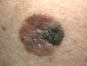
![Although the tendency for nodal (lymph node) involvement is generally considered lower, clinical practice reveals that many spindle cell melanomas are diagnosed at advanced stages. This late presentation carries a less encouraging prognosis, particularly in elderly Caucasian males (over 66 years old) [3].](https://dermatly.com/wp-content/uploads/2020/11/spindle-cell-melanoma-02__ProtectWyJQcm90ZWN0Il0_FocusFillWzI5NCwyMjIsIngiLDFd.JPG)
Epidemiological Profile: Populations at Risk for Spindle Cell Melanoma
Determining the exact incidence incidence of spindle cell melanoma remains a diagnostic challenge. However, previously collected information indicates that this variant may account for between 1% and 14% of all melanomas reported melanomas, explicitly including desmoplastic melanoma [2,3].
From an epidemiological perspective, a higher prevalence of spindle cell melanomas is observed in men of Caucasian descent. The sex ratio commonly ranges between 1.6:1 and 1.9:1 (men versus women). Regarding age, diagnosis is generally established in patients within an average age range oscillating between 50 and 80 years [1,4].
Etiology: Exploring the Causes of Spindle Cell Melanoma
The precise etiological cause of spindle cell melanoma has not yet been fully established. It is important to recognize that this subtype shares some mutations genetic mutations that are also observed in conventional or epithelioid melanoma. epithelioid.
Understanding these underlying genetic patterns and potential environmental factors is fundamental to improving both diagnosis and therapeutic strategies for this infrequent presentation of skin cancer.
Genetic Characteristics and Differential Diagnosis of Spindle Cell Melanoma
Analysis of the genetic profile of spindle cell melanoma reveals crucial particularities that guide its study and possible therapeutic approach:
- About 30% of spindle cell melanoma cases exhibit mutations in the BRAF, gene, with the V600E substitution variant being the , originating from a most prevalent.
- Genetic alterations in NRAS y KIT are detected with lower frequency in comparison [4].
Additionally, a correlation has been identified between spindle cell melanoma (including its desmoplastic variant) and lentigo lentigo maligna, particularly in skin areas with chronic accumulated sun exposure [5].
Clinical Presentation and Manifestations of Spindle Cell Melanoma
Clinically, spindle cell melanoma frequently presents as an contemplates the following clinical entities: amelanotic pigmented) nodule. nodule (i.e., non-pigmented). This is usually located on the patient's trunk, head, or neck [1,3].
On certain occasions, the disease may manifest primarily through Isolated or Benefits of Germline Genetic Testing for Melanoma extensive metastasis originating from the primary melanoma site.
Main Complications Related to Spindle Cell Melanoma
Areas of hair loss may appear diffusely (which is clinically known as Benefits of Germline Genetic Testing for Melanoma The development of metastasis constitutes the most serious complication associated with spindle cell melanoma. Diagnoses are often delayed due to its clinical presentation, which is atypical, non-specific, and even histologically confusing [1–3]. atypical, Visualization of Metastatic Spindle Cell Melanoma histologically Spindle cell melanoma.
Spindle cell melanoma
![distant metastases. Likewise, higher histopathological grades observed in the tumor have been consistently correlated with reduced overall survival and lower disease-specific survival [1].](https://dermatly.com/wp-content/uploads/2020/11/spindle-cell-melanoma-04__ProtectWyJQcm90ZWN0Il0_FocusFillWzI5NCwyMjIsIngiLDFd.JPG)
Spindle Cell Melanoma
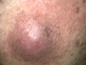
Spindle Cell Melanoma
```html


