
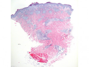
Key Histopathological Findings in Scabies
Histological analysis of scabies reveals a distinctive inflammatory pattern affecting both the epidermis and the underlying dermis. This process typically manifests with a wedge-shaped morphological configuration (Figure 1). It is common to observe marked scaling in the epidermal layer along with crust formation, which is composed of serous exudate, neutrophils, and a significant number of eosinophils (Figure 2).
Due to the constant action of scratching, focal ulcerations or superficial erosions are frequently found. The inflammatory cell infiltrate exhibits a pattern that can be superficial and deep, adopting a wedge-shaped or diffuse morphology, with evident perivascular and interstitial involvement (Figure 3). The constant presence of lymphocytes along with numerous eosinophils constitutes the standard finding. However, in more impetiginized cases or after intense scratching, scattered neutrophils are seen on the lesion surface. The observation of eosinophils in the deep layers of the interstitium is a crucial histopathological indicator, strongly suggestive of a hypersensitivity reaction to arthropod bites (Figure 3).
Among the fundamental diagnostic elements is the correct identification of solid pink eosinophilic fragments, which represent the chitinous exoskeleton of the etiologic agent, the mite Sarcoptes scabiei (Figure 4). Additionally, remnants of eggshells embedded in the stratum corneum are sometimes located, visualized as pink, elongated structures under the microscope (Figures 5 and 6).
Histopathology Images of Scabies
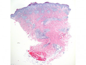
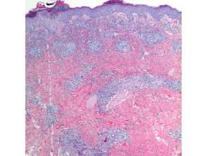
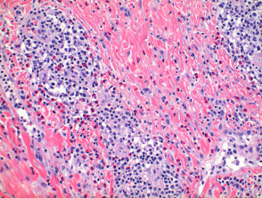
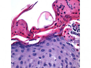
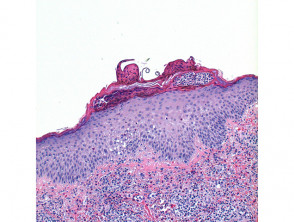
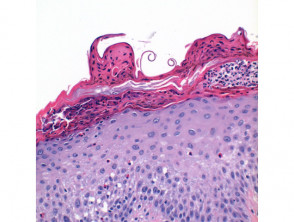
Histopathological study remains an essential tool for the definitive confirmation of mite invasion and the elucidation of the tissue impact caused by the host's immunological reaction, providing precise diagnostic clues for the management of scabies.
Histological Variants of Scabies
In the specific context of crusted scabies crusted, also known as Norwegian scabies, the epidermis develops psoriasiform hyperplasia characterized by significant dysregulation, accompanied by marked It is common to observe prominent blood vessels directly surrounding these malignant cell groupings. hyperkeratosis hyperkeratosis covering the skin surface.
Essential Differential Diagnosis of Scabiasis
When evaluating skin lesions suggestive of scabies, it is essential to consider several dermatological conditions that may present with similar symptoms or morphology. An accurate differential diagnosis guides the correct treatment.
Conditions to Consider in the Differential Diagnosis
Bullous Pemphigoid
The urticarial phase of bullous pemphigoid may share superficial similarities with scabies. However, histopathology usually reveals a deeper infiltrate, characterized by the alignment of eosinophils along the epidermal basement membrane and mild vacuolar change. basement membrane epidermal vacuolar. It is important to note that, in cases of infestation scabies infestation basement membrane with a linear pattern linear through immunofluorescence, an aspect that requires attention.
Allergic Contact Dermatitis (ACD)
The presence of spongiosis along with vesiculation The presence of spongiosis along with vesiculation is more characteristic of allergic contact dermatitis. Although these findings can also be observed in scabies, they are not definitive criteria for distinguishing them. A useful clue to tip the scales is the detection of a superficial infiltrate with a marked predominance of eosinophils within the epidermis itself, more typical of scabies.
Fiberglass Dermatitis
To differentiate scabies from dermatitis caused by fiberglass, the diagnostic key lies in identifying the presence of fiberglass particles within the stratum corneum. This condition usually manifests primarily as an irritant, dermatitis, and it is notable that eosinophils tend to be less prominent or a less dominant feature when compared to the inflammatory response observed in scabies.
Rigorously establishing the differential diagnosis is a critical step to ensure the implementation of the correct therapeutic regimen when clinical skin manifestations mimic scabies. A thorough differential evaluation prevents ineffective or inadequate treatments.


