
Radiological Imaging: An Essential Tool in Oncological Diagnosis
Radiographic studies produce detailed visuals of soft tissues and underlying bone structures. This category encompasses various imaging modalities utilizing radiation or magnetic fields, including computed tomography (CT Scan), CT imaging, ultrasound scans, and magnetic resonance imaging (MRI).
Key Radiological Images in Melanoma Assessment
The Role of Radiographic Studies in Melanoma Management
For patients diagnosed with melanoma, radiographic investigations offer significant benefits, aiding in the early detection of metastases or providing crucial reassurance. However, these methods inherently carry risks, such as identifying benign lesions (false positives) or failing to detect existing metastatic disease (false negatives).
Clinical follow-up must integrate symptomatic evaluation and physical examination of the primary tumor site, alongside regional lymph nodes. Furthermore, routine self-skin examinations remain indispensable, as imaging techniques are seldom effective at identifying new **primary melanomas** (primary melanomas).
There are three primary justifications for utilizing radiographic studies in melanoma patients:
- Initial patient staging, which determines if the cancer poses a high risk and has already spread to internal organs.
- Assessing the efficacy of treatments previously administered, such as targeted therapies and immunotherapies [1–3].
- Continuous monitoring for patients at high risk of recurrence or progression following a resection, aiming to quickly detect any reappearance of the disease [1].
Clinical guidelines stipulate that imaging studies are not recommended for melanoma in situ (Stage 0) or for thin, non-aggressive (Stage I or II) melanomas, provided the patient exhibits no clinical signs or symptoms of metastasis. Consequently, advanced staging or prophylactic surveillance is reserved for individuals facing a substantial likelihood of developing distant metastases. [4].
While primary tumors and localized nodal metastases are typically identifiable through clinical examination, monitoring distant organs
Visceral or metastatic malignant melanoma visceral o metastatic is often asymptomatic asymptomatic until advanced stages, at which point it can be very difficult to eradicate surgically [1].
Radiographic Imaging Used in Staging and Surveillance of Malignant Melanoma
Genetic testing radiological images Basic radiological tests such as chest X-ray (CXR) can identify previously undetected metastatic melanoma; however, their two-dimensional soft tissue visualization capabilities are limited [5]. For this reason, most patients are referred for Computed Tomography (CT), Positron Emission Tomography-CT (PET scans.-CT), ultrasound scans, and Magnetic Resonance Imaging (MRI).
Use of CT and PET-CT in Advanced Oncology
CT and PET-CT are essential tools in the staging and follow-up of many solid malignant neoplasms. solid.
- CT offers detailed three-dimensional views of body anatomy.
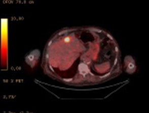
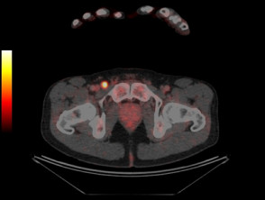
- PET-CT incorporates a fluorodeoxyglucose (FDG) tracer, which highlights regions with high metabolic activity, a common characteristic in solid malignant tumors, including melanoma.
- PET-CT complements CT by detecting metabolically active disease in sites such as lymph nodes, which might appear structurally normal on a standard CT scan.
- While PET-CT locates metastases with high accuracy in most locations, it is not the ideal modality for the brain, due to the high background metabolic activity in that area, with diagnostic CT or MRI being superior [6]. more [6].
Ultrasound Imaging for Regional Lymph Node Surveillance
Ultrasound is used specifically to monitor regional lymph nodes areas looking for possible metastases. It is important to note that the accuracy of ultrasound results is intrinsically nuclear dependent on the skill and experience of the evaluating sonographer [7,8].
Magnetic Resonance Imaging (MRI) for High Diagnostic Resolution
MRI provides images with exceptionally high resolution, making it valuable for certain diagnoses:
- It is the preferred method for searching for metastatic disease within the brain.
- Furthermore, MRI can be used to investigate in greater detail those areas that have been flagged as suspicious on a PET-CT study, before proceeding to surgery or biopsy [9].
Criteria for Radiographic Surveillance in High-Risk Melanoma Patients
Periodic radiographic evaluations are appropriate for patients diagnosed with stage III malignant melanoma, particularly those with histologically confirmed nodal dissemination, or presence of in-transit or satellite metastases. These groups face a considerable risk of experiencing recurrence or progression to stage IV metastatic melanoma [10–12]. Likewise, the relatively poor prognosis associated with internal organ and distant lymph node involvement becomes considerably more complex with purely physical methods. Therefore, active surveillance using advanced techniques becomes indispensable in these critical scenarios. histologically, Periodic radiographic evaluations are appropriate for patients diagnosed with stage III malignant melanoma, particularly those with histologically confirmed nodal dissemination, or presence of in-transit or satellite metastases. These groups face a considerable risk of experiencing recurrence or progression to stage IV metastatic melanoma [10–12]. Likewise, the relatively poor prognosis associated with internal organ and distant lymph node involvement becomes considerably more complex with purely physical methods. Therefore, active surveillance using advanced techniques becomes indispensable in these critical scenarios.
Active Surveillance and Imaging Protocols for Advanced Stage Melanoma
Consideration of an active surveillance protocol is justified for stage IIC melanoma [12].
- There is notable inconsistency in international guidelines regarding the frequency and type of imaging surveillance in melanoma patients, as these resolutions are based more on expert consensus than on rigorous clinical evidence.
- Comparing findings from published research studies is difficult since they have included patient cohorts with very disparate melanoma stages.
- Until recently, the scarcity of effective systemic treatment systemic options limited the interest in strictly evaluating the utility of radiographic investigations for follow-up.
Implementing a personalized surveillance protocol is fundamental for these high-risk patients, as this ensures early detection of any possible disease progression, thus allowing for the timely initiation of appropriate treatments.
- Follow-up using radiographic studies has undergone significant evolution thanks to the development development of immunotherapies and targeted therapies.
- The US National Comprehensive Cancer Network (NCCN) recommends performing computed tomography (CT) or PET-CT every 3 to 12 months for asymptomatic patients with stage IIB-IV melanoma [13].
- In contrast, the European Society for Medical Oncology (ESMO) suggests only a physical examination every three months [14].
Ultrasound surveillance of the nodal nodal region is considered appropriate in the following clinical scenarios:
- Monitoring for recurrence in those patients whose sentinel node was positive but who did not receive a complete nodal dissection.
- Surveillance in individuals who did not undergo a sentinel node biopsy but who still present a high risk of progression to stage III disease due to unfavorable primary tumor characteristics.
- For initial staging of the nodal region; however, it must be recognized that the accuracy of ultrasound is lower than that obtained through sentinel node biopsy [15–17].
In melanomas classified as stage I or II, ultrasound surveillance proves superior to regional clinical examination of the lymph nodes, although the actual benefit in long-term survival derived from this ultrasound follow-up remains uncertain [8]. Only a small group of patients may benefit from early detection of nodal metastases, or from avoiding unnecessary surgeries, while another portion ends up undergoing surgical procedures that could have been prevented due to initially suspicious findings [8].
Key Benefits of Radiographic Investigations in Melanoma Surveillance
Metastases detected in isolation through surveillance or staging by imaging can potentially be treated with surgical resection, radiotherapy or systemic therapies [18–20]. Patients receiving the most modern therapies exhibit notably higher survival rates [21–23].
- According to data collected in Australia, Positron Emission Tomography (PET) managed to identify metastases up to six months earlier than other routine exams, whether physical or imaging [24].
- Two research studies indicated that the therapeutic approach for patients with stage III melanoma was modified between 19% and 35% after performing PET-CT studies [25–27].
- Both PET-CT and conventional CT have the ability to identify the presence of additional primary neoplasms [28–29].
Additional Advantages When Selecting Imaging Modalities
- Ultrasound constitutes a non-invasive procedure that does not involve radiation exposure.
- Unlike computed tomography scans, Magnetic Resonance Imaging (MRI) does not emit ionizing radiation.
The selection of the most appropriate surveillance method must be carefully weighed, considering the patient's individual risk profile and the balance between early detection and the burden of frequent radiological exams. Continuous research seeks to standardize these protocols to optimize clinical outcomes in the management of advanced melanoma, especially now that more effective systemic treatments exist to control metastatic disease.
Challenges and Frequency of Radiographic Surveillance in Melanoma
The lack of rigorous randomized clinical trials prevents definitively confirming whether routine imaging surveillance programs lead to a significant improvement in the survival of melanoma patients.
It is crucial to highlight that, despite the results of the scans, the detection of the majority of melanoma recurrences—whether local or distant—falls to the patients themselves or their partners. This is because these findings are often easily visible or simply palpable palpable on the skin surface or in the nodal region [30,31].
- The ability of imaging techniques to discover distant metastases in asymptomatic patients shows great variability, ranging between 15% and 72%, depending on both the initial stage and the type of study used [31–33].
- Positron Emission Tomography (PET-CT) rarely manages to identify metastatic disease smaller than 5 mm [34–36].
- The benefit derived from using PET-CT to discover occult metastases in patients with a positive sentinel lymph node proved to be marginal, reporting rates between 0.5% and 3.7% [37–40].
Additionally, surveillance using imaging techniques carries the inherent risk of identifying nonspecific lesions or producing false-positive results, which can significantly increase patient anxiety and motivate the need for unnecessary medical interventions.
This culminates in the need to investigate results suggesting false metastases [1,41].
- When inconclusive or false-positive findings are detected, it becomes essential to perform additional investigations, often including invasive procedures [41,42].
- PET surveillance in patients diagnosed with metastatic stage IIIA melanoma has revealed a false-positive rate fluctuating between 7% and 14% [1,43,44].
- In one of these investigations, 86% of these false-positive reports led to subsequent biopsies [1].
Several studies have evaluated the overall accuracy of PET-CT for both initial staging and continuous patient follow-up.
- PET-CT sensitivity and specificity were documented at 65% and 99% for regional disease detection, 86% and 91% for distant metastases, and 80% and 87% for overall staging, respectively [45].
- The ultrasound: This tool is useful for diagnosing chronic Ultrasound showed the highest performance in sensitivity (60%) and specificity (97%) for regional nodal staging, with values of 96% and 99% respectively for directly involved lymph nodes [45].
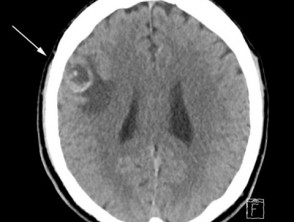
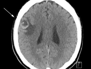
- The reliability of both PET and cranial CT in identifying brain metastases is limited [46,47]. In a group of almost 700 patients with metastatic melanoma, cutaneous it was determined that 12% had asymptomatic brain metastases detected by computed tomography [48].
- Cerebral magnetic resonance imaging (MRI) offers a superior evaluation for intracranial metastatic disease, intracranial, however, it is contraindicated. contraindicated for patients carrying metallic implants.
- A single Positron Emission Tomography-Computed Tomography (PET-CT) detected 24% of recurrences in 110 asymptomatic patients diagnosed with stage IIB to IIIB melanoma [44].
- In a subgroup of 170 patients undergoing surveillance with PET-CT imaging, melanoma recurrence was detected in 38% of cases, with 69% of these recurrences being asymptomatic. A negative PET-CT at 18 months demonstrated negative predictive values between 80% and 84% regarding the absence of recurrence during the full follow-up of 47 months (mean) [1]. Of the patients whose recurrence recurrence.
was found by PET-CT, 33 (52%) underwent resection with curative potential, although only 16% remained disease-free after 24 months [1].
Determining the Optimal Frequency for Melanoma Surveillance Studies.
- To date, neither the ideal periodicity nor the most advisable total duration for implementing radiographic surveillance studies has been established with certainty.
- Routine clinical practice recommends administering positron emission tomography every 6 months as a surveillance method for patients classified in the high-risk group [1,23].
It is consistently observed that the highest incidence of melanoma recurrences typically occurs during the first two or three years following the initial diagnosis of the disease [1,11].


