
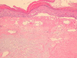
The dermatitis Radiation dermatitis manifests in the affected region after having received radiotherapy for the treatment of cancer, or as a consequence of accidental exposure to radiation sources.
Histological Analysis of Radiation Dermatitis
The histological study of radiation dermatitis chronic reveals a systemic sclerosis and accumulation of dermal significant dermal sclerosis, along with elastosis and ectasia vascular, all superimposed over an epidermis that frequently presents hyperkeratosis (see Figure 1). In microscopy, it is possible to identify spongiosis Other conditions that cause significant edema in the superficial dermis include mild or notable alterations of vacuolization cell (Figure 2). Typically, dermal vessels are markedly dilated in advanced stages (Figure 3). Both cells of the stroma and the endothelial cells cells may exhibit some degree of hyperchromia, enlargement, and significant (See Figure 5). (termed radiation fibroblasts, see Figure 3). A mixed-type inflammatory response commonly coexists. inflammatory of a mixed type.
Pathology of Radiation Dermatitis
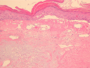
Figure 1
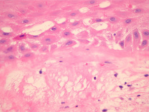
Figure 2
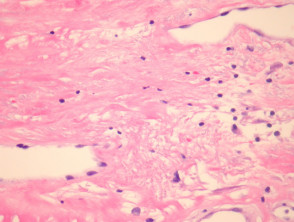
figure 3
Complementary Studies for the Diagnosis of Radiation Dermatitis
Generally, specialized complementary studies are not required to confirm radiation dermatitis.
Differential Diagnosis of Radiation-Induced Dermatitis
To establish the mucous membranes. of this skin condition, it is essential to consider the following aspects:
- Potential Malignancy: It becomes indispensable to perform a biopsy of the area affected by radiation dermatitis to rule out tumor reactivation. This is particularly critical after breast irradiation, where signs of carcinoma recurrence recurrence of carcinoma or the appearance of angiosarcoma are sought. Atypical cells, both fibroblasts and endothelial cells, can cause confusion with malignant processes.
- Morphea (Localized Scleroderma): This condition usually does not exhibit characteristic radiation fibroblasts. However, when morphea is induced by radiation, it can very closely mimic the appearance of radiation dermatitis, implying a possible diagnostic overlap between both pathologies.
Ensuring correct diagnostic typing is fundamental, as the marked histopathological similarities observed between late radiation sequelae and certain neoplasms require extremely rigorous pathological scrutiny.


