
Understanding Pyogenic Granuloma: Definition and Etiology
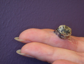
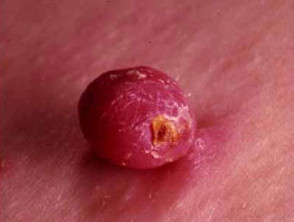
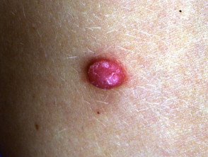
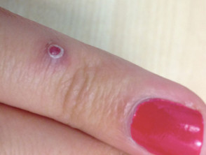
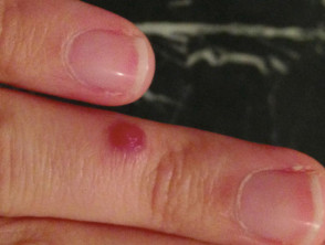
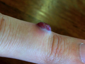
The **pyogenic granuloma** is a relatively common condition characterized by a reactive proliferation of capillary blood vessels. Clinically, it manifests as a bright red nodule whose surface resembles ground meat or a raspberry. Despite its benign nature, pyogenic granulomas can cause significant discomfort and present with profuse bleeding upon minor trauma.
This diagnosis is also known by several synonyms, including lobular capillary hemangioma, *pyogenicum* granuloma, and telangiectatic granuloma.
Clinical Images of Pyogenic Granuloma

What Causes Pyogenic Granuloma? Etiological Factors
The exact etiology of pyogenic granuloma remains unknown. However, various factors have been identified as possible contributors to its development:
- Trauma: Certain cases emerge at the site of a recent minor lesion or wound, such as a small puncture.
- Infection: The presence of *Staphylococcus aureus* has frequently been detected in the involved lesion.
- Hormonal Influences: They can manifest in up to 5% of pregnancies and occasionally correlate with the use of oral contraceptives.
- Drug Induction: Multiple lesions can appear in patients receiving oral retinoids (such as acitretin or isotretinoin) or protease inhibitors.
- Viral Infection: While a theoretical possibility, its role in pathogenesis has not been proven.
Epidemiology and Risk Factors for Pyogenic Granuloma
Who is most prone to developing Pyogenic Granuloma?
Pyogenic granuloma can manifest in individuals of any ethnic background. However, incidence is notably higher in women, frequently associated with hormonal changes during pregnancy. Although uncommon in infants under six months, this condition is regularly observed in both children and young adults.
Clinical Characteristics: Signs and Symptoms of the Growth
Typically, the initial pyogenic granuloma presents as a painless macule with a color ranging from bright red, reddish-brown, or bluish-black. This growth exhibits rapid proliferation over a few days or weeks, reaching an average final size of 1 to 2 cm, although exceptionally it can reach up to 5 cm. A distinctive feature is its tendency to bleed easily upon minor trauma; occasionally, it may ulcerate, forming a scab painful crust. Generally, a solitary lesion develops, although rare reports of cases with multiple growths exist.
The most frequent anatomical locations include the scalp, neck, upper trunk, hands (particularly the fingers), and feet. When the pyogenic granuloma is related to gestation, it is more common to observe it on the mucosal surfaces of the mucosa lip or inside the oral cavity.
Diagnostic Process for Pyogenic Granuloma
Clinical Determination of Diagnosis
The diagnosis of pyogenic granuloma is, in most cases, clinical, based on its characteristic presentation and evolution.
Confirmation via histology histology is crucial, especially when there is diagnostic uncertainty regarding malignant skin lesions, such as amelanotic melanoma, which are part of the differential diagnosis for skin cancer. contemplates the following clinical entities: melanoma, skin cancer mucous membranes. of the cancer On histopathological examination, pyogenic granuloma reveals a lobulated proliferation of blood vessels immersed in inflammatory tissue.
Treatment Options for Pyogenic Granuloma Lesions
Some pyogenic granulomas have the potential to resolve spontaneously, which is observed more frequently in those linked to pregnancy. If the underlying cause is drug ingestion, the lesion tends to disappear upon discontinuing that medication.
There are several established procedures for the removal of pyogenic granuloma:
- Surgical excision. surgical excision.
- Curettage and cauterization: This involves scraping the lesion with a curette and then cauterizing the feeding blood vessel to minimize the probability of recurrence. blood vessel feeding blood vessel to minimize the probability of recurrence.
- Laser surgery laser to remove the bump and seal the base, or the use of pulsed dye laser to reduce the size of small lesions.
- The cryotherapy Cryotherapy.
- Chemical cauterization using silver nitrate proves effective in small lesions.
- The efficacy of lotion Imiquimod cream.
- Experimentally, early use of topical 1% propranolol reactions. ointment has shown promising results in the pediatric treatment of pyogenic granulomas [1].
Recurrence after treatment is a common event because the blood vessels irrigating the lesion extend in a conical shape deep into the dermis. dermis. In these persistent scenarios, the most definitive and effective removal method is the complete excision of the affected area (excision), followed by closure with sutures.


