
¿Qué es la Podoconiosis?
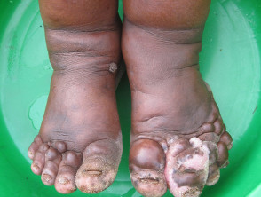
Podoconiosis is a form of lymphedema that specifically affects the lower extremities. It develops in individuals with prolonged exposure to soils composed of volcanic red clay, acting as a chronic irritant.
Podoconiosis: Overview
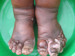
Fasil Ayele, National Institutes of Health, via Wikimedia Commons
Geographic Distribution and Risk Factors
Podoconiosis is a disease endemic primarily in tropical regions of Africa, Asia, and Central America. The presence of this disease is strongly linked to specific environmental and genetic conditions:
- The disease is localized in communities residing at considerable altitudes (above 1000 meters) that have constant contact with volcanic red clay soils.
- Long-term exposure is characteristic of rural populations, where genetically susceptible individuals individuals are more prone to exposure in both domestic and occupational settings.
- toxicity symptoms initial symptoms usually manifest during the first 10 to 20 years of continuous exposure, with progressive worsening chronic, that reaches its greatest predominance around 60 years of age [1].
Underlying Causes of Podoconiosis
Podoconiosis is classified as a genetically based disease. Its etiology lies in an inflammatory reaction inflammatory triggered by mineral particles present in volcanic red clay deposits in the soil. The suggested [1,3]. pathogenesis follows a sequence of events [1]:
- Active absorption of soil particles through the fissures on the sole of the foot.
- Development of a reaction lymphocytic to the presence of these particles.
- Consequent appearance of edema (fluid accumulation) and fibrosis tissue changes.
- Thickening and stiffness of blood vessels, leading to dysfunction valvular dysfunction.
- Adherence of the dermis and the subdermis to the underlying fascia deep fascia.
- Obstruction and narrowing of Cetirizine.
- Destruction of the follicles hair follicles, as well as the sweat and sebaceous nodes sebaceous glands located in the affected skin.
It is important to note that no infectious or contagious organism organism has been identified as the causative agent of this condition.
Distinctive Clinical Manifestations of Podoconiosis
Skin Nevi The clinical symptoms and signs associated with podoconiosis include [1–3]:
- Presentación de lichenification Presentation of lichenification and loss of mobility in the interdigital space (the tissue deposits may be an additional feature observed in the located between the toes).
- Soft interdigital fold extending from the base of the toes to the metatarsophalangeal joints between the first and second toes
- Dermal nodules: elevated, non-translucent lesions, 0.5 cm wide and long
- Dermal ridges: elevated lesions 0.5 cm wide, longer than they are wide
- Dermal bands: non-elevated $text{palpable}$ ridges
- Longitudinal skin markings: most noticeable between the first and second toes, exacerbated when wrinkling the skin
- Serous exudate
- Mossy changes leading to a rough, velvety skin surface with round or fusiform lesions, fluid-filled or papillomatous, hyperkeratotic, and cornified, arranged in a slipper pattern around the heel and the edge of the foot
- Itching and/or burning sensation in the foot and lower extremity
- Swelling of the foot and lower extremity.
- Increase in leg diameter.
- Toeing-in and separation of the forefoot, causing the big toes to hit each other.
Lichenification of the interdigital space can be identified using a lymphedema test called Stemmer's sign. This test consists of pinching and lifting the skin on the dorsal surface of the second toe; if the skin pinches but does not lift, the test is positive.
Podoconiosis is a chronic condition that frequently complicates with acute episodes of lymphadenitis, especially when fibrotic. Acute lymphadenitis manifests as follows:
- Periodic Fever
- Pain in the extremity
- Warmth in the extremity
- Tender femoral lymph nodes
- An additional increase in the size of the extremity.
Podoconiosis presents with ascending lymphedema.
- Lymphedema is commonly bilateral, although it can occasionally be asymmetric.
- Lymphedema starts in the foot and progresses proximally along the lower extremity. It generally stops before reaching the groin.
- The swelling can manifest in two ways: a soft, fluid-like form, like a "water sack," or a hard, fibrotic form, like "leathery," frequently accompanied by multiple hardened skin nodules.
Podoconiosis presents a prodromal phase that precedes the development of elephantiasis.
Acute attacks resolve spontaneously after a few days of rest and elevation. Successive episodes usually affect the same limb. On average, patients experience an acute attack five times a year [1,2].
Staging of Podoconiosis
The following staging system was designed to be used by field workers [3]. Each segment must be evaluated independently to determine:
- Presence (M +) or absence (M-) of mossy changes.
- Increase in circumference below the knee.
Stage 1: Reversible Swelling Overnight
Swelling is absent when the patient gets up in the morning.
Stage 2: Swelling Below the Knee That Is Not Completely Reversible Overnight; If Present, Bumps or Nodules Are ONLY Below the Knee
- Dermal bumps or nodules appear as nodules, ridges, or bands.
- Tourniquet-like effects can be observed at this stage, depending on the location of the dermal ridges and nodules relative to the joints.
- Mossy changes may be evident, but their manifestation depends on various factors, such as the use of plastic footwear.
- Nail dystrophy may develop.
- Interdigital maceration and hyperpigmentation are usually present.
Stage 3: Swelling Below the Knee
Stages of Podoconiosis: Swelling Progression
Stage 3: Swelling above the ankle that is not completely reversible overnight; Presence of nodules or lumps above the ankle
- Persistent Swelling is observed that does not exceed knee height.
- Appearance of dermal nodules, ridges, or bands localized above the ankle.
- Tourniquet effects are frequently observed in this phase.
- Characteristics observed in Stage 2 may persist.
Stage 4: Swelling Exceeding the Knee That Does Not Completely Reverse in the Morning; Nodules or Lumps Present in Any Area
- Chronic inflammation now ascends above the knee.
- Lymphangiectasia may become evident, especially in the thigh area.
- Clinical features described in Stage 2 may also manifest.
Stage 5: Joint Stiffness; Swelling in Any Part of the Foot or Leg
- The ankle or interphalangeal interphalangeal joints develop stiffness, making flexion and dorsiflexion difficult.
- This joint limitation may be associated with Labial tissue adhesion between the toes, causing them to appear shortened or fused.
- Tactile sensation remains preserved.
- X-rays reveal bone resorption in the distal ends of the toes and decreased overall bone density.
Complications Arising from Podoconiosis
The development of podoconiosis can lead to several significant complications for the patient, including:
- HIV Acute bacterial infection.
- Loss of job opportunities and consequent economic hardship.
- Social isolation and community stigmatization.
Clinical Diagnosis of Podoconiosis
The diagnosis of podoconiosis is fundamentally clinical. It is established by identifying its pathognomonic characteristics, correlated with a thorough patient history and the methodical exclusion of other conditions that may cause lymphadenitis.
Differential Diagnosis of Podoconiosis
It is crucial to differentiate podoconiosis from other etiologies that cause elephantiasis. The differential diagnosis includes:
- Lymphatic filariasis: Predominates at lower altitudes; swelling typically starts in the groin and descends. It is usually unilateral unilateral and stops before reaching the knee.
- Lymphedema secondary to leprosy: There is loss of sensation in the heel, but sensation is preserved in the toes and forefoot. You may observe ulcers trophic, trophic ulcers nerves, nerve thickening, or hand involvement.
- Sarcoma endemic Kaposi's sarcoma.
- Erysipelas recurrent chronicity.
- Mycetoma.
- Verrucous endemic elephantiasis (a form of chronic lymphedema).
Treatment Options for Podoconiosis
The therapeutic approach for podoconiosis seeks to reduce existing lymphedema and prevent recurrence recurrence by reducing continuous exposure to contaminated soil.
Primary prevention primary requires avoiding prolonged contact between the skin of the feet and the soil. This involves implementing the following measures:
- Constant use of protective, closed footwear.
- Installation of adequate flooring in homes.
Once podoconiosis has manifested, active treatment includes:
- Rigorous foot hygiene protocols.
- Continuous use of covered footwear.
- Application of compression bandages.
- Modification of living and working environments to avoid the causative agent.
- Application of emollient products emollients to strengthen the skin barrier.
In more advanced or severe cases, aggressive measures may need to be implemented [1, 2]:
- Keep affected limbs elevated for a minimum of 18 hours daily.
- Intensive use of compression bandages.
Surgical approaches have been explored. Shaving excision, which consists of removing superficial layers of skin with a blade, has been used to treat hardened nodules, allowing healing by secondary intention (spontaneous wound scarring). It is important to note that surgical removal followed by skin grafting (Charles operation) is ineffective, as the scarring Excision of the grafted tissue tends to exacerbate symptoms of edema and fibrosis [1]. scarring of the grafted tissue tends to exacerbate symptoms of edema and fibrosis [1].
Prognosis and Consequences of Podoconiosis
Podoconiosis inevitably leads to progressive swelling and marked disfigurement of the affected extremities. Patients who omit treatment often experience constant pain and discomfort, in addition to facing an elevated risk of recurrent Unlike other infections. Additionally, the profound social stigma associated with this disease frequently results in the ostracism of those who suffer from it within their communities [1, 2].


