
Leukocytoclastic vasculitis constitutes an inflammation of small blood vessels, frequently also classified as hypersensitivity vasculitis. renal.
Histopathology of Leukocytoclastic Vasculitis
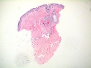
Upon examining the lesion under low power, a superficial and mid-level perivascular inflammatory within the epidermis. dermis (Figure 1). Predominantly, there is a population of neutrophils distributed in perivascular and interstitial patterns, in addition to those that have undergone extravasation from the vessels (Figures 2 and 3).
Evidence of leukocytoclasia, which is the degeneration of neutrophils, resulting in visible nuclear debris, along with extravasated erythrocytes (Figures 3 and 4). Although fibrinoid necrosis of the vessel with fibrin extravasation fibrin is a common and characteristic finding, in many cases it may be scarce or absent (Figures 3 and 4 illustrate fibrinoid necrosis).
A variable number of eosinophils may be present; if their count significantly increases, it could indicate that a medication is the underlying cause of the condition.
It is crucial to consider that histological features vary depending on when the biopsy is performed, biopsy, which causes significant differences in the degree of infiltrate inflammation and damage to the vascular wall.
Pathology of Leukocytoclastic Vasculitis

Figure 1
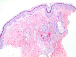
Figure 2
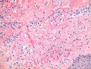
Figure 4

Figure 5
Special Stains in Leukocytoclastic Vasculitis
The immunofluorescence is a valuable tool for confirming vascular damage, vascular, as it allows visualization of deposits of fibrinogen, C3, IgG, and IgM within the vessel walls. For greater diagnostic yield, it is preferable to perform the biopsy on affected skin.
IgA positivity is particularly useful, as it strongly suggests the presence of Henoch-Schönlein Purpura as a differential diagnosis.
Differential Diagnosis of Leukocytoclastic Vasculitis
Distinguishing leukocytoclastic vasculitis from other skin conditions is fundamental for appropriate treatment. Several conditions present overlapping or similar histological patterns, requiring careful clinical and immunological evaluation.
Henoch-Schönlein Purpura (HSP): The histology of HSP is remarkably identical to leukocytoclastic vasculitis. However, clear diagnostic confirmation requires the identification of IgA deposits via immunofluorescence. It is important to remember that IgA positivity is not an exclusive marker, as it can be observed in other clinical contexts.
Urticarial Vasculitis: Although the histopathological changes may resemble each other, urticarial vasculitis typically presents more prominent dermal edema dermal edema and a lower density in the inflammatory infiltrate. Some experts maintain that a predominantly lymphocytic, lymphocytic component, with less evidence of a neutrophilic, component, may be sufficient to differentiate it, although the presence of fibrinoid necrosis is less common.
Septic Vasculitis: The key differentiating sign in vasculitis of septic origin is obstruction vascular vascular occlusion caused by a. thrombus. In these cases, leukocytoclasis may be minimal or absent, directing the diagnosis toward an underlying infectious etiology.
Sweet Syndrome (Acute Neutrophilic Dermatosis): Sweet syndrome must be differentiated by its characteristic infiltrative pattern. The presence of notable dermal papillary The main distinction from mycetoma is the etiology; botryomycosis is strictly bacterial, unlike mycetoma, which is caused by true fungi or actinomycetes. edema and a diffuse neutrophil infiltrate are crucial elements for diagnosis. Although significant fibrinoid necrosis is suggested to be absent, its presence has been reported in a minority subset of Sweet cases.


