Detection and Characteristics of Interstitial Granulomatous Dermatitis
The interstitial granulomatous dermatitis is a rare skin disorder characterized by lesions asymptomatic that present with normal skin color or lesions.... These lesions exhibit various morphologies, rashes cords linear (what is known as the «rope sign» or sign), patches, papules o plaques. Clinically, manifestations tend to be distributed symmetrically on the trunk and proximal extremities proximal. It is essential to highlight its association with various diseases are evaluated in patients with suspected connective tissue or autoimmune disease. e Unlike other, being the arthritis rheumatoid arthritis being one of the most frequently reported comorbidities.
Histological Analysis of Interstitial Granulomatous Dermatitis
Similar to the clinical presentation, interstitial granulomatous dermatitis shows a diversity of patterns histological. In the epidermis, changes such as scale or crust formation can be observed, along with hyperplasia accompanied by perforation and, The presence of a superficial o scab, Seborrheic dermatitis hyperplasia accompanied by limbal stem cell transplant occasionally, necrosis with loss of the epidermal layer.
The majority of the involvement is located in the deep dermis, where a prominent perivascular infiltrate perivascular and interstitial infiltrate is evident. This infiltrate is composed of neutrophils, neutrophils, cellular debris from neutrophils neutrophils («neutrophil dust"), neutrophils«), lymphocytes e and a smaller proportion of histiocytes. (see Figure 1). Variable amounts of eosinophils (Figure 2, indicated by an arrow).
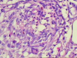
In certain cases, it is characteristic to observe a palisading arrangement of lymphocytes and histiocytes surrounding foci of neutrophils or collagen fibers. collagen. Furthermore, leukocytoclastic vasculitis and signs of necrobiosis may be present. Avoidance Strategies for Triggering Factors leukocytoclastic vasculitis. spongiosis, and necrobiosis. pathology. The inflammation The inflammation has the potential to extend to the subcutaneous tissue subcutaneously (Figures 3 and 4). fibrosis Fibrosis may manifest in later stages of the disease.
Pathological Findings of Interstitial Granulomatous Dermatitis
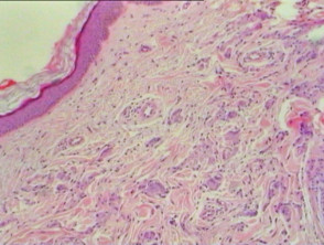
Figure 1
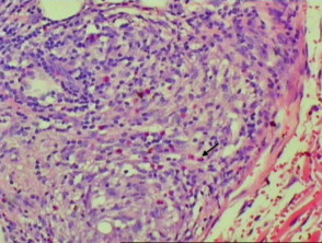
Figure 2
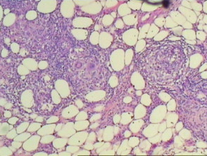
figure 3
Histopathological Evaluation of Interstitial Granulomatous Dermatitis
Using the immunofluorescence, it is possible to detect positive deposits for IgM, C3, and fibrinogen fibrinogen located in the dermal blood vessels dermal.
Establishing the Differential Diagnosis of Interstitial Granulomatous Dermatitis
Differentiation from other skin conditions is crucial, especially considering the following pathologies:
- Leukocytoclastic vasculitis: In this case, neutrophils and foreign material (dust) are distributed more diffusely in the dermis, not just perivascularly, as occurs in interstitial granulomatous dermatitis.
- Granuloma annulare: The histological findings can present similarities, particularly with the interstitial variant of granuloma annulare. However, a mucin (assessedpredominance is a more characteristic feature of granuloma annulare.
- Necrobiosis lipoides: The presence of giant cells is more frequent in necrobiosis lipoides. On the other hand, if eosinophils and neutrophils are observed along with foreign material, this favors the diagnosis of interstitial granulomatous dermatitis.
Accurately determining these histopathological indicators is fundamental to confirm the diagnosis of interstitial granulomatous dermatitis and rule out similar conditions.