
Understanding Favre-Racouchot Syndrome: Clinical and Histological Findings
The discovery of nodular elastosis accompanied by cysts and comedones, conditions that correlate with skin aging, chronic solar radiation, and smoking, defines syndrome Favre-Racouchot syndrome.
Histological Analysis of Favre-Racouchot Syndrome
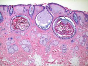
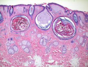
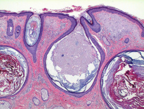
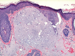
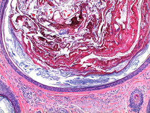
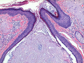
At low magnification, the histology of Favre-Racouchot syndrome reveals the presence of dilated and plugged comedones in the hair hair follicles, along with the formation of cysts changes epidermal cysts (references in Figures 1 and 2). This picture is established over an epidermis epidermis nodules that shows severe solar damage, which forms notable nodules in the superficial dermis (Figure 3). The comedones are densely packed with compact keratinous material, while the epidermal cysts contain looser laminated keratin (Figures 4 and 5).
Histological Illustrations of Favre-Racouchot Syndrome
Histological Variants of Favre-Racouchot Syndrome
Although the classic presentation of Favre-Racouchot syndrome includes significant sun damage along with the presence of cysts and comedones, there are case reports that manifest these lesions without marked sun exposure. This suggests that constant solar radiation may not be the indispensable pathogenic factor in all manifestations of this condition.
Differential Diagnosis Considerations
It is crucial to distinguish Favre-Racouchot syndrome from other skin conditions. Morphologically similar or even identical changes can be observed in patients with chloracne or severe forms of acne vulgaris presenting with severely photoaged skin. However, in these alternative diagnoses, the prominence of cyst formation tends to be less. Therefore, a thorough and precise clinical correlation is essential to establish the correct diagnosis.
A detailed understanding of the histopathological findings, as described in the figures, is essential to differentiate Favre-Racouchot syndrome from sun-induced dermatoses. While the etiology is firmly linked to chronic photoaging, the consistency in clinical presentations helps guide appropriate treatment.


