

What is Carcinoma In Situ (CIS) of the Oral Cavity?
Oral carcinoma in situ (CIS), also known as oral intraepithelial carcinoma or squamous cell carcinoma in situ, represents a precancerous stage where malignant cells remain strictly confined to the epithelium. This condition is clearly distinguished from invasive squamous cell carcinoma (SCC), where cells have begun to penetrate beyond that superficial layer.
Risk Factors for Oral Carcinoma In Situ
Oral intraepithelial carcinoma affects approximately 0.5% of the global population, although this figure varies considerably based on gender, ethnicity, and geographic region.
There is a very strong correlation with tobacco use (being six times more prevalent in smokers than in non-smokers) and alcohol consumption, regardless of the pattern or type of alcoholic beverage. Additionally, it has been associated with areca nut chewing and oral submucous fibrosis.
Generally, this pathology is diagnosed in adulthood, and its incidence gradually increases over the years.
- Less than 1% of males under 30 years old present with oral carcinoma in situ.
- Up to 8% of men over 70 years old and 2% of women over 70 years old are diagnosed with oral cavity carcinoma in situ.
- It is rarely observed before age 30, reaching its peak incidence after age 50.
- It primarily affects middle-aged and elderly men.
- Non-smoking individuals tend to develop the condition at an older age.
Exploring the Clinical Manifestations of Oral Carcinoma In Situ
In its initial phases, oral carcinoma in situ frequently presents as a well-demarcated, grayish or whitish plaque that is slightly elevated or subtly merges with the surrounding mucosa. The condition can appear as a single focal lesion or present multifocally and extensively.
Clinically, two main patterns of presentation can be identified:
Homogeneous Form: Uniform Appearance
This variant is characterized by coloration and texture that remain consistent throughout the lesion:
- It presents a uniform white color (in stages prior to diagnosis, this might have been labeled as leukoplakia).
- Its surface is smooth, flat, and maintains a constant thickness.
The surface of these lesions may feel leathery, smooth, rough, wrinkled, or present superficial fissures. Generally, this clinical manifestation is painless.
Non-Homogeneous Form: Irregularity in Color and Texture
This form is defined by variations in pigment or surface morphology:
- It is predominantly white, or a mixture of white and red (which might have previously been termed erythroleukoplakia).
- Its surface is irregular, potentially appearing flat, nodular, exophytic (outward growth), or verrucous.
Subtypes have been identified within the non-homogeneous form, including nodular, verrucous (including proliferative verrucous), and speckled lesions. This clinical presentation may be associated with mild discomfort or localized pain.
The most common location for the development of oral carcinoma in situ is the inner cheek area (buccal mucosa), followed in frequency by the following areas:
- The gums (alveolar mucosa).
- The lower lip.
- The floor of the mouth (sublingual region).
- The lateral or ventral surface of the tongue.
- The soft palate.
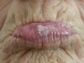
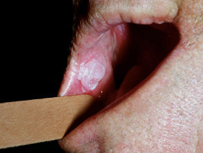
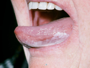
Association of Oral Leukoplakia with Squamous Cell Carcinoma (SCC)
A significant proportion of oral carcinomas are linked to a prior history of long-standing carcinoma in situ, particularly in the proliferative verrucous variant.
During the initial stages of cancer, the appearance or symptoms may be subtle or nonexistent. The development development of carcinoma manifests through more evident clinical changes, including ulceration, hardening or induration, easy bleeding, and the formation of a tumorgrowth or excrescence.
Key Risk Factors in the Development of SCC Associated with Leukoplakia
- Trichothiodystrophy Dysplasia atypicalatypical changes at the histological level) is considered the most significant predictive factor. However, it is crucial to note that dysplastic lesions may show spontaneous resolution, and lesions without apparent dysplasia can progress to cancer.
- Anatomical location is key: the floor of the mouth (under the tongue) and the lateral/inferior surfaces of the tongue present a higher risk.
- Clinical type: non-homogeneous speckled verrucous leukoplakia, particularly the proliferative variant, increases risk.
- Female sex has been noted as a risk factor in certain studies.
- When the preceding carcinoma in situ is not related to tobacco use.
- The prolonged duration of the cellular alteration (carcinoma in situ).
- The large size of the diagnosed lesion.
- The presence of the bacterium Candida albicans. Candida albicans. It is important to consider that this coexistence is more frequently observed in lesions located in the oral commissures or on the dorsal surface of the tongue, sites that are statistically less common for cancer development.
To date, no molecular tumor markers molecularhave been identified that can reliably predict cancerous evolution in a specific patient or lesion. The exact role of human papillomavirus (HPV) in this process is still under investigation.
Guide for the Confirmatory Diagnosis of Oral Leukoplakia
Establishing an accurate diagnosis requires specific procedures to evaluate the intrinsic risk of malignancy of the lesion.
- The biopsy Biopsy.
- of clinically suspicious oral leukoplakia is a mandatory step. This allows ruling out other pathologies and determining the presence and degree of epithelial dysplasia. It is recommended to wait two weeks after the initial presentation. This period allows for evaluating the clinical response to initial treatments aimed at contributing factors, such as treating Candida infections, modifying oral hygiene techniques, or achieving tobacco cessation. cessation cessation.
- The biopsy can be incisional incisional local anesthetic or excisional (complete), and can be performed singly or multiply. The technique is chosen under biopsies local.
- anesthesia hardenedor general, depending on the site, the number of biopsies.
- needed, and the selected method. Samples should prioritize symptomatic areas or, if the lesion is asymptomatic, those presenting with red coloration or.
The histopathology indurated Epithelialareas. limb Clinical correlation is not always sufficient to predict the presence of dysplasia, carcinoma in situ, or invasive carcinoma in the final histopathological analysis.
Histopathology.
of oral leukoplakia often does not offer a conclusive diagnosis by itself. The epithelial changes observed cover a spectrum ranging from hyperplasia atrophy hyperkeratosis. to the pathology loss of normal architecture of the cell layers. If there is any suspicion of high risk, referral to a specialist and rigorous follow-up are essential for the management of oral leukoplakia and the prevention of squamous cell carcinoma. It can present with.
hyperplasia
(a thickening of the tissue) and manifest along with It is essential to remember that HNC exhibits a considerable rate of hyperkeratosis.
- . Dysplasia, which involves atypical cellular alterations, is graded as mild, moderate, severe, carcinoma in situ, or invasive carcinoma. It is essential that the pathology report clearly details the presence and degree of severity of any detected dysplasia.
- Surgical excision. surgical excision.
- Management of Oral Carcinoma In Situ (CIS) laser There is still no certainty as to whether early and active treatment of oral squamous cell carcinoma in situ prevents its progression to an invasive form. It should be noted that the recurrence.
- of the lesion after receiving treatment is a considerably high probability. Topical Immediate elimination of associated harmful habits, such as smoking. photodynamic.
Surgical excision.
- Use of vaporization or CO2 laser as an excision method.
- There are complementary alternatives such as the administration of retinoids (isotretinoin or acitretin) and the application of photodynamic therapy.
Regardless of whether the lesion has been treated or not, it is imperative to establish a continuous, lifelong follow-up regimen: mucosa Schedule clinical evaluations every 3 to 12 months.


