
Onychomycosis, also known as tinea unguium, is defined as a fungal infection that affects the nail bed, matrix, or plate, predominantly caused by dermatophytes. It is important to note that Candida-type fungi and non-dermatophytes are usually more prevalent in regions with tropical climates.
Histology and Evaluation of Onychomycosis
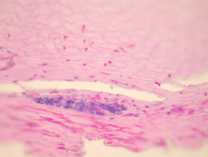
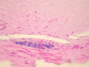
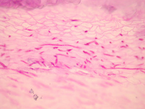
To evaluate the presence of fungal forms in onychomycosis, it is essential to examine sections of the affected nail using specialized stains such as PAS or GMS. Often, nail samples reveal the presence of exudate and accumulations of neutrophils, neutrophils, along with fungi that have penetrated the nail tissue (see Figure 1). Specifically, the PAS stain in Figure 2 allows visualization of dermatophytes invading the nail plate.
Pathology of Onychomycosis
Key Special Studies for the Diagnosis of Onychomycosis
PAS or GMS stains are fundamental diagnostic tools, as they allow for clear identification of the organism organisms that have managed to penetrate the nail tissue.
Differential Diagnosis of Onychomycosis in Pathology
The mucous membranes. S100 protein. pathology requires ruling out other conditions that mimic fungal infection.
A key point is colonization by non-dermatophytes: these organisms can establish themselves in the subungual space. However, unless there is clear evidence that they have actively penetrated the nail plate, they are not attributed as the cause of onychomycosis.
Regarding psoriasis, although neutrophilic aggregations can be observed (forming neutrophilic (forming accumulations), PAS/GMS stains will be negative for the identification of fungal organisms.
In cases of pitted keratolysis, the bacterial cause can be confirmed by Gram stain, where thin and filamentous bacterial forms will be evident, differentiating them from fungal structures. bacterial thin and filamentous, differentiating them from fungal structures.


