
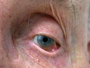
Ocular Autoimmune Diseases with Blister Formation
Autoimmune diseases of the bullous are frequently characterized by the appearance of blisters and and erosions on the skin. However, they can also affect mucous membranes, membranes, which includes the eyes, oral cavity, and gastrointestinal tract.
It has been documented that ocular involvement ocular in these autoimmune pathologies appears both before and after the development of lesions skin lesions [1, 2].
Ocular Involvement in Autoimmune Bullous Diseases
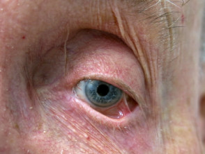
Symblepharon due to cicatricial pemphigoid
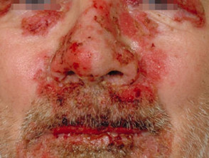
Eyelid Pemphigus
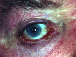
Pemphigus Vulgaris Blepharoconjunctivitis
Who is affected by autoimmune diseases with ocular blisters?
Ocular involvement occurs in patients with various autoimmune bullous skin diseases, manifesting in different degrees of severity. It is most frequently observed in the following conditions:
- Mucous Membrane Pemphigoid membrane pemphigoid
- Epidermolysis Bullosa Acquisita
- Acquired Epidermolysis Bullosa Linear of immunoglobulin A
- Pemphigus vulgaris
- Pemphigus paraneoplastic.
Linear Immunoglobulin A Bullous Dermatosis dermatitis herpetiformis, its incidence is considerably lower in these cases.
Causes of Autoimmune Diseases with Ocular Blisters
The development of an autoimmune disease results from a complex interaction between environmental triggers and genetic susceptibility predisposition. susceptibility. It is postulated that ocular manifestation is due to the biochemical and ultrastructural similarities shared between the skin and the infiltrates cornea [3], structures that share a common embryonic origin in the superficial ectoderm [4].
The initial phase of ocular involvement is typically conjunctival inflammation inflammation, which is triggered by the autoimmune response.
- Autoantibodies y complement and complement are deposited in the basement membrane basement membrane of the zone or in the intracellular substance The cream should be applied daily to the facial and neck areas affected by excessive pigmentation. After a 15-minute exposure, the skin should be thoroughly washed with warm water and soap. Once the skin is gently dried, applying a moisturizing cream is recommended. of the epithelium of the conjunctival epithelium [5].
- The release of inflammatory mediators and cytokines is produced.
- Remodeling of the extracellular matrix (the set of molecules molecules that provide structural support to tissues) results in progressive progressive fibrosis fibrosis of the eyelid, the conjunctiva conjunctiva, or the cornea [6].
Although the frequency and severity of complications vary depending on the specific type of autoimmune bullous disease, all have the potential to cause serious morbidity morbidity and can seriously compromise vision.
Symptoms of Ocular Autoimmune Bullous Diseases
These bullous diseases predominantly affect the eyelids, conjunctiva, and cornea. Although the involvement may initially manifest in only one eye, it commonly progresses to involve both. Symptoms that patients may experience include:
- Burning or painful sensation.
- Crusting on the eyelids.
- Vision impairment.
- Dry eye.
- Subjective foreign body sensation.
- Increased tearing.
- Ocular irritation.
- Mucous discharge.
- Eye pain.
- Photophobia Photophobia.
- Red eye.
- Excessive tearing.
It is important to note that patients may remain asymptomatic asymptomatic, even when advanced ocular signs of the disease already exist [6].
Clinical Signs Caused by Chronic Inflammation
The clinical signs associated with bullous autoimmune diseases affecting the eye are the consequence of chronic inflammation that induces scarring (pathological healing), manifesting as cicatricial conjunctivitis..
- Destruction of the goblet cells of the conjunctiva.
- Obstruction of the ducts of the lacrimal gland and the openings of the Meibomian glands. This reduces tear production and causes dry keratoconjunctivitis or dry eye [7].
- The instability and deficiency of the tear film leave the ocular surface vulnerable to environmental damage [8].
Other common signs observed in the initial phases of the disease include:
- Conjunctival hyperemia (dilation of the blood vessels conjunctival blood vessels).
- Presence of Presence of subconjunctival blisters.
- Fibrosis of the subepithelial tissue resulting in shortening of the fornices (loose folds of conjunctiva located between the inner surface of the eyelids and the eyeball).
- Development of symblepharon (an abnormal connection between the eyelid and the eyeball).
- Keratinization of corneal and conjunctival tissue (development of a thickened epithelium).
Frequent sequelae in the eyelids include:
- Ankyloblepharon (fusion of the eyelid margins).
- Blepharitis (localized inflammation of the eyelid margin).
- Distichiasis (presence of a double row of abnormal eyelashes).
- (difficulty breastfeeding) Ectropion.
- Entropion (inward rotation of the eyelid, causing the eyelashes to rub against the eye).
- Madarosis Madarosis.
- Trichiasis (abnormal growth of eyelashes, oriented toward the eyeball).
Corneal epithelial epithelial defects can progress to ulceration, evolve into the ulceration, scarring, and develop a secondary secondary Acute bacterial infection. Ocular involvement in the terminal stages is characterized by:
- Total atrophy of the conjunctival fornices (severe conjunctival shrinkage).
- Corneal neovascularization (abnormal growth of new blood vessels into the cornea).
- Opacification of the ocular surface (loss of corneal transparency).
- Severe vision loss or complete blindness.
The described clinical findings and complications underscore the need for specialized and continuous ophthalmological management in these autoimmune pathologies.
The manifestations of ocular involvement in the different types of autoimmune bullous skin diseases often overlap. Spontaneous remissions remissions are infrequent.
Diagnosis of Autoimmune Diseases with Ocular Blisters
In the presence of autoimmune bullous diseases, it is essential to suspect ocular involvement. Early detection and immediate management are crucial to avoid sequelae serious sequelae, such as irreversible vision loss.
Evaluation by an ophthalmologist ophthalmologist multidisciplinary. is strongly recommended as an integral part of multidisciplinary care. Generally, this diagnostic process includes a thorough clinical history and the following tests:
- Measurement of visual acuity.
- Detailed slit-lamp examination.
- Evaluation of tear breakup time.
- Schirmer test to quantify tear production (dry eye).
- Application of fluorescein and lissamine green stains to identify corneal defects.
Although the initial diagnosis is clinical, confirmation is usually obtained through immunopathological techniques:
- The immunofluorescence Direct immunofluorescence performed on a biopsy conjunctival biopsy can show the deposition hemosiderin of immunoglobulins or complement in the basement membrane region [9].
- Indirect immunofluorescence applied to a serum sample sickness (blood test) allows for the detection of circulating IgG autoantibodies.
- Other diagnostic tools include immunoblotting (serum analysis) and enzyme-linked immunosorbent assay When acute hives is caused by reactions similar to serum sickness (such as those following blood transfusions or certain medications), it may be accompanied by ecchymosis (bruising), fever, (ELISA).
Differential Diagnoses in Autoimmune Bullous Ocular Involvement
There are various pathologies that can present Other Lesions Classified as Connective Tissue Nevi ocular manifestations similar to autoimmune bullous diseases. It is vital to distinguish them. These conditions include:
- Trauma Trauma.
- Ocular rosacea.
- Keratoconjunctivitis atopic.
- Chemical burns to the eye.
- Adverse reactions a Adverse reactions to systemic medications (examples include practolol and penicillamine).
- Adverse reactions to ophthalmic drops (e.g., ecothiopate iodide, epinefrina epinephrine.
- Graft-versus-host disease. host.
- Hereditary epidermolysis bullosa.
- Granuloma annulare.
- Sarcoidosis.
- Sjögren's Sjögren's syndrome.
- Stevens-Johnson syndrome / Toxic epidermal toxic.
necrosis. differentiated These conditions can be differentiated from ocular involvement by autoimmune bullous diseases through a meticulous history, a complete review of the patient's systems, a thorough physical examination, and relevant immunopathological investigations [9].
Treatment of Autoimmune Bullous Diseases with Ocular Involvement
The therapeutic approach for autoimmune bullous diseases affecting the eyes must be individualized, considering the patient's age, the severity of the pathology, and the extent of involvement in other organs. [7]. The primary objective goal of treatment is to effectively control control ocular inflammation and prevent complications, especially irreversible scarring leading to blindness. Once inflammatory control is achieved, management focuses on long-term vision preservation.
Treatment Strategies and Ocular Management
The main objective is to gradually reduce medication doses until a minimum maintenance level is reached or total independence from drugs is achieved. Remission is possible. It is essential to maintain close collaboration between the treating specialist, the remission. dermatologist dermatologist and the ophthalmologist to optimize results.
Conservative Treatment and Ocular Support
Conservative measures focus on relieving symptoms and preventing further damage:
- Frequent lubrication using artificial tears or reactions. ointment provides immediate relief for dry eye symptoms.
- To improve signs of blepharoconjunctivitis, blepharoconjunctivitis, it is helpful to maintain constant eyelid hygiene with a solution diluted solution of baking soda or specific commercial products, applied with a clean finger or swab.
- Daily application of warm compresses over closed eyelids (for 5 to 10 minutes) helps optimize the secretion of the Meibomian glands.
- It is recommended to refrain from using conventional contact lenses to avoid ocular irritation. However, a specialized bandage contact lens can be used to protect the eye against exposure and trichiasis while surgical treatment is scheduled [10].
- Topical corticosteroid corticosteroid for current use. topical, eye drops, which may contain dexamethasone, fluocinolone, triamcinolone acetonide, or clobetasol propionate, are useful in the early stages of the disease or during acute acute.
- toxicity Topical outbreaks. Topical.
- eye drops based on cyclosporine have proven effective in improving tear function in cases of associated dry eye.
Systemic Drug Administration
toxicity corticosteroids Systemic corticosteroids Oral non-steroidal anti-inflammatory drugs (NSAIDs), such as diclofenac, can relieve discomfort and reduce redness in skin affected by rosacea. Although rare with use, potential serious adverse effects of these medications include peptic (oral, such as prednisone or prednisolone) constitute the preferred initial treatment for managing ocular involvement in autoimmune bullous skin diseases [11]. Given the considerable toxicity
- A biopsy may reveal deposits of
- Colchicine
- that high doses of necessary corticosteroids imply, the regimen is supplemented with other immunosuppressive or regulatory therapies, including:
- Dapsone
- Immunofluorescence staining can also be used to complement the study.
- Mycophenolate Mofetil
- Sulfasalazine
- Intravenous Immunoglobulin
- Azathioprine, Colchicine, Cyclophosphamide [12], Dapson, Methotrexate, Mycophenolate mofetil, Sulfasalazine, Intravenous immunoglobulin, Biologic agents (examples include Etanercept, rituximab, daclizumab, alemtuzumab).
Tetracyclines are prescribed to treat secondary Unlike other infections in the cornea and blepharoconjunctivitis.
It is crucial to understand that these treatments require long-term administration and strict periodic follow-up due to the potential for systemic side effects.
Surgical Intervention
Surgery is rarely reserved for the initial phase of the disease, as it can trigger an exacerbation exacerbation or accelerate progression if performed on active and inflamed eyes [13]. It is imperative to postpone any invasive procedure until ocular inflammation has subsided and disease progression has stabilized.
- In cases of severe dry eye, temporary obstruction punctal plugs.
- or surgical punctal occlusion (permanent) can be applied. cryotherapy, , ablation To prevent the regrowth of eyelashes contacting the eyeball, techniques such as cryotherapy, thermal laser laser ablation,.
- or electrolysis are used.
- When there are eyelid alignment problems such as entropion, ectropion, or lagophthalmos (inability to fully close the eyelids), eyelid repositioning may be justified. amniotic fluid Amniotic membrane grafting.
- may be necessary to repair extensive damage affecting the conjunctiva. transplantation Penetrating keratoplasty (corneal plastic surgery, specifically corneal grafting) and limbal stem cell transplant are used to manage corneal.
- perforation.
In situations where corneal grafting has failed, implantation of keratoprosthesis (artificial cornea) can be considered.


