
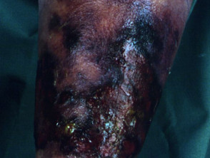
Understanding Necrotizing Fasciitis: A Severe Infection
The necrotizing fasciitis is an extremely serious medical condition, characterized by a rapid and destructive bacterial infection that attacks the soft tissue and the fascia, the fibrous sheath that covers the muscles. Within these layers, the bacteria multiply rapidly, secreting potent toxins y muscle enzymes. This process leads to in the vessel wall. (clotting) and subsequent obstruction of the blood vessels, resulting in necrosis, or death, of the soft tissues and surrounding fascia.
The main types of necrotizing fasciitis are classified according to the causative microorganisms and their routes of infection:
- Type I: It is polymicrobial, implying the involvement of several bacterial strains simultaneously.
- Type II: It is specifically caused by group A hemolytic streptococcus (streptococcus) or staphylococci, including methicillin-resistant strains (MRSA).
- Type III: It involves gangrene bacterial gas gangrene, usually caused by Clostridium.
- Other Types: Include infections by marine organisms (such as Vibrio o Aeromonas hydrophila, species, sometimes categorized as Type III) and Unlike other fungal infections (such as Candida and zygomycetes, sometimes classified as Type IV).
Specific Typologies of Necrotizing Fasciitis
Necrotizing Fasciitis Type I
The bacteria that cause Type I necrotizing fasciitis are varied and include Staphylococcus aureus, Haemophilus, Vibrio and a mixture of aerobic y anaerobic, strains, such as Escherichia coli y and. Bacteroides fragilis. This form typically manifests in older populations or in patients with significant comorbidities such as diabetes.
Necrotizing Fasciitis Type II
This variant has received considerable media attention and is popularly known as the «flesh-eating disease.» Type II can affect any age group, and it is notable because it can also thrive and infect individuals who are otherwise considered completely healthy.
Necrotizing Fasciitis Type III (Gas Gangrene)
Type III necrotizing fasciitis is primarily caused by Clostridia perfringens or, less frequently, by Clostridia septicum. This infection frequently develops after a severe injury or major surgery. Its distinctive characteristic is the production of gas under the skin, which generates a crackling sound audible to the touch known as crepitus. Furthermore, intravenous drug users who inject «black tar» heroin» subcutaneously are at risk of developing this form of clostridial necrotizing fasciitis.
Infections Caused by Other Organisms
Necrotizing fasciitis resulting from marine organisms is commonly associated with wound contamination by seawater, cuts sustained from contact with fish fins or spines, or the ingestion of raw shellfish. These cases are more prevalent in patients with pre-existing liver conditions. If not treated rapidly, these marine infections can be extremely dangerous and fatal in less than 48 hours.
On the other hand, mycotic necrotizing fasciitis tends to complicate traumatic wounds in immunocompromised.
There are other clinical terms used to describe various presentations of necrotizing fasciitis, including hemolytic streptococcal gangrene, Meleney's ulcer ulcer and accumulation of dermal acute, acute dermal gangrene, hospital-acquired gangrene, suppurative fasciitis and synergistic necrotizing cellulitis.
When the condition affects the perineal, genital, and perianal, perianal regions , it is called. Fournier's Gangrene. This specific presentation carries a particularly high mortality rate, which can range between 15% and 50% of cases.
How Does Necrotizing Fasciitis Develop? Who Is at Risk?
Anyone can contract necrotizing fasciitis; in fact, it is a surprising fact that nearly half of documented cases of streptococcal necrotizing fasciitis have occurred in young and previously healthy individuals. The disease requires the convergence of certain conditions to establish itself, which generally include:
- A breach or opening in the skin that provides an entry route for bacteria into the body. This can result from minor injuries, such as a small cut, scrape, puncture, or injection, or it can arise after major trauma trauma or a surgical procedure (such as laparoscopy,
- Recent invasive procedures (e.g., sclerotherapy, endoscopic gastrostomy , thoracostomy, C-section, hysterectomy). Sometimes, no entry point is identified., Cervicofacial necrotizing fasciitis can manifest after a jaw fracture or a dental infection.
- Direct contact with a carrier of the bacteria, or when the bacteria is already present in another body region of the individual.
- Especially invasive bacterial strains, such as streptococci, which manage to evade the immune system and release a toxin.
- invasive toxin, called cysteine toxin protease SpeB, capable of dissolving tissue. In the pediatric population, Type II necrotizing fasciitis can be a complication of chickenpox. Other etiologies in children include omphalitis, necrotizing enterocolitis, and urachal anomalies.
- Risk factors associated with necrotizing fasciitis include:.
Use of aspirin and non-steroidal anti-inflammatory drugs.
- advanced age and inflammatory.
- Diabetes.
- State of.
- immunosuppression Obesity..
- Substance abuse.
- Severe.
- chronic Malignancy. serious.
- Detailed Pathophysiology of Necrotizing Fasciitis.
The infectious process begins in the superficial fascia. Enzymes and
released by the responsible microorganisms trigger the proteins necrosis necrosis of the fascial layers. This horizontal spread of the infection is often not superficially visible on the skin, which can lead to a delay in diagnosis. Subsequently, the infection advances vertically toward the dermis and deeper structures. The formation of thrombi occludes the arteries arteries and veins, culminating in ischemia.
and subsequent tissue death.
- Specifically, streptococcal strains secrete: cytokines M proteins, which cause an intense inflammatory response with the release of multiple cytokines TNF(such as IL-1, IL-6, and.
- TNFα). Exotoxins neutrophils, that have the ability to destroy neutrophils, which facilitates bacterial proliferation and tissue destruction.
Furthermore, both aerobic and anaerobic bacteria produce gases—such as hydrogen, nitrogen, hydrogen sulfide, and methane—that degrade hyaluronic acid, thus facilitating the rapid spread of the infection.
Clinical Manifestations: Signs and Symptoms of Necrotizing Fasciitis
While clinical manifestations vary among patients, most experience a combination of the following signs.
Initial Prodromal Symptoms
- The most frequent location of the infection is the lower extremity. However, necrotizing fasciitis can also affect the upper limbs, the perineum, the buttocks, the trunk, as well as the head and neck.
- Generally, symptoms manifest within 24 hours following a minor skin injury.
- The associated pain is usually extremely severe at the initial presentation and tends to intensify progressively.
- Systemic flu-like symptoms may occur, including nausea, fever, fever malaise.
- , diarrhea, dizziness, and a generalized feeling of malaise. As the body experiences dehydration, intense thirst develops.
Subsequent Symptoms
Clinical Evolution of Necrotizing Fasciitis: Days 3 to 5
As necrotizing fasciitis progresses, its manifestations rapidly become more severe:
- The affected area begins to swell noticeably and may take on a purplish hue, along with the appearance of a rash.
- rash.
- . Extensive dark markings develop, evolving into blisters filled with dark fluid.
- The edema The tissue begins to die, manifesting as blackening (necrosis).
- Significant.
- edema is a common feature. A fine crackling sensation or *crepitus* is experienced under the skin, indicating the presence of trapped gas in the tissues. nerves The unbearable pain persists until necrosis or gangrene destroys the peripheral nerves.
- and the pain subsides. The infection may show resistance to treatment, with no apparent improvement after antibiotic administration.
Between days 4 and 5, the patient's condition deteriorates critically: they present with high fever and dangerously low blood pressure. The infection has spread to the bloodstream, leading the body into a state of toxic shock. In this advanced phase, the patient may show altered levels of consciousness or fall into a coma.
Metastatic abscesses may arise in vital organs such as the liver, lungs, spleen, brain, pericardium, and, rarely, the skin.
Illustrative Images of Necrotizing Fasciitis
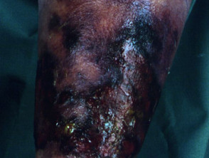
Necrotizing Fasciitis
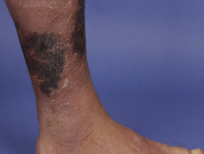
Necrotizing Fasciitis
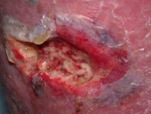
Necrotizing Fasciitis
Review more detailed images of necrotizing fasciitis to better understand its severity.
Diagnosis of Necrotizing Fasciitis: Clinical Evaluation
A thorough clinical history and physical examination are essential for diagnosing necrotizing fasciitis. Extreme caution is required when evaluating immunocompromised patients, as their symptoms may present atypically. atypical.
- Infections caused by *Clostridium* and *Streptococcus*, stemming from traumatic or surgical wounds, tend to manifest more rapidly compared to those caused by other microorganisms.
- The The finger test (or skin-peeling test) is highly pathognomonic. A 2 cm vertical subcutaneously is made in the injured skin, and the index finger is inserted into the tissue. The test is positive if the finger passes through the.
- subcutaneous.
- tissue without encountering resistance. When separating the tissue during the incision, little to no adherence to the underlying fascia is observed. Necrotic o tissue orpus drains from the fascial planes..
- Clear fluid, similar to dishwater, often seeps through the skin.
- A relevant sign is that necrotizing fasciitis typically presents with minimal or no bleeding in the surgical area.
Laboratory Analysis for Confirmation
Laboratory results usually reveal the following patterns:
- Leukocyte count greater than 15.4 x 10⁹/L.
- Serum sodium concentration <135 mmol/L.
- Elevated CRP CRP (C-Reactive Protein) levels (> 16 mg/dL).
- Elevated CK (Creatine Kinase) level (> 600 U/L).
- Urea level greater than 18 mg/dL.
Additionally, blood culture culture biopsy , deep tissue organismbiopsy.
, and Gram stain are essential to identify the causative organism(s) and guide the optimal selection of antibiotics. If *Staphylococcus* is detected...
Diagnosis and Management of Necrotizing Fasciitis Staphylococcus aureus, If infection by.
*Staphylococcus aureus*.
, a sensitivity test should be performed to detect MRSA. Generally, blood cultures are negative for *Clostridium* species.
In immunocompromised patients or those who have suffered severe trauma, fungal culture is essential.Imaging Tools for Diagnosis, Techniques such as X-ray, computed tomography ( CT , translated here as Connecticut due to the original tag) and inflammation Magnetic Resonance.
Imaging (MRI) are crucial for identifying fluid accumulation, inflammation, and the presence of gas within the soft tissues.
Laboratory Risk Indicator (LRINEC) for Necrotizing Fasciitis.
The Laboratory Risk Indicator for Necrotizing Fasciitis (LRINEC) is a tool designed to differentiate NF from other tissue infections by evaluating six analytical parameters. A score equal to or greater than 6 strongly suggests the presence of necrotizing fasciitis. However, it is important to note that this test is not universally applicable and its absolute reliability is not guaranteed.
Detailed LRINEC Parameters
- The score is calculated based on the following laboratory values:
CRP (mg/L) ≥ 150: 4 points3Leukocyte count (× 103):
- 3
- / mm
- 3):
< 15: 0 points
- 15-25: 1 point
- > 25: 2 points
- Hemoglobin (g/dL):
> 13.5: 0 points
- 11-13.5: 1 point
< 11: 2 points
- 11-13.5: 1 point
Sodium (mmol/L) < 135: 2 points
- Creatinine (µmol/L) > 141:
2 points
Glucose.
- (mmol/L) > 10:.
- 1 point.
- What is the Fundamental Treatment for Necrotizing Fasciitis? Once necrotizing fasciitis has been diagnosed, it is imperative to initiate therapeutic treatment immediately and aggressively..
- The patient requires hospitalization, frequently in an Intensive Care Unit (ICU).
- The causative microorganisms must be identified and treated with high-dose intravenous antibiotics. Initial empirical therapy usually includes penicillin, clindamycin, metronidazole, cephalosporins, carbapenems, vancomycin, and linezolid. Drug selection is adjusted once culture results are received. It is absolutely vital that an experienced surgeon urgently proceeds with the removal of all necrotic tissue through e immunoglobulin debridement.
. Supplemental oxygen, intravenous fluids, and supportive medication may be necessary to maintain stable blood pressure. excision. Hyperbaric.
oxygen therapy and intravenous immunoglobulin may also be considered. ulcer Immediate surgical debridement is the most important factor in improving the survival rate and preventing serious complications associated with necrotizing fasciitis. All infected tissue must be removed by excision. It is common for repeated surgical debridements to be required over several days until complete control of the infection is achieved.
Once the acute phase of the infection has subsided, the wound must be closed, using skin grafts if necessary. Vacuum-assisted wound closure systems can be valuable tools to facilitate the healing of a
persistent.
ulcer.
Prognosis and Expected Outcome of Necrotizing Fasciitis ulceration, sepsis The determining factor for reducing the risk of mortality and severe disfigurement in cases of necrotizing fasciitis is early diagnosis and the speed of the treatment implemented.


