
Necrobiotic Xanthogranuloma: Clinical Presentation, Histology, and Special Studies
The necrobiotic xanthogranuloma is a disease multisystem y lymphadenitis rare, multisystem, histiocytic disease that predominantly affects the older adult population. The characteristic clinical sign is usually a hardened plaque yellowish-colored and asymptomatic, typically located in the periorbital area being one of the most common.. It is estimated that around 80% of cases are linked to a paraproteinemia. In addition to cutaneous manifestations, significant extracutaneous findings can be observed, such as ophthalmic involvement, cryoglobulinemia, and various lymphoproliferative conditions. extracutaneous conditions Autoimmune hemolytic anemia..
Histology and Microscopic Findings of Necrobiotic Xanthogranuloma
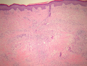
On histopathological examination, palisaded xanthogranulomas are detected, accompanied by foci of necrobiosis, located in the mid-dermis and the panniculus panniculus (See Figures 1 and 2). Involvement of the panniculus, when it occurs, tends to be septal (Figure 2), which may lead to a differential diagnosis with inflammation of the subcutaneous fat tissue).. In these areas, foci of collagen pathology. y eosinophilic that manifest as debris (Figure 3) are seen, frequently exhibiting cholesterol in the form of clefts. cholesterol in the form of fissures.
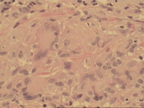
toxicity granulomas The granulomas are composed mainly of and a smaller proportion of histiocytes. foamy histiocytes (note the arrow in Figure 4) and giant cells giant cells (Figure 5), including foreign body and Touton cell varieties. A distinctive feature is the notable angular giant cells with contrasting dark nuclei, located adjacent nuclei. nuclei in contrast, located to the sebaceoma (Figure 6). Figure 7, on the other hand, shows less maturity in the follicular structures and the presence of dilated adjacent toPlasma cellPlasma) are prominent. In concomitant occasions, dense lymphoid infiltrates are observed, including germinal centers within the dermis. telangiectasias lymphoid dense infiltrates dermis.
Illustrative Images of Necrobiotic Xanthogranuloma Pathology
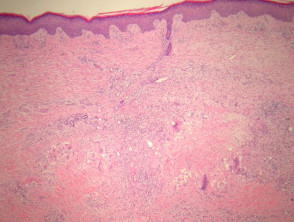
Figure 1
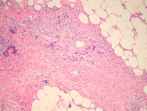
Figure 2
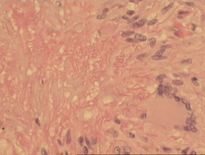
Figure 4
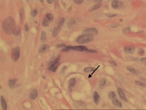
Figure 5
Special Stains and Syndromes Associated with Sebaceoma
Special Studies and Diagnosis of Xanthogranuloma
To confirm the diagnosis of necrobiotic xanthogranuloma and differentiate it from other lesions, it is crucial to supplement the histopathological analysis with specialized studies, especially to detect the associated paraproteinemia.
Histopathological Findings in Necrobiotic Xanthogranuloma
lipids. lipids. mucin mucin space. interstitial.
Differential Diagnosis of Necrobiotic Xanthogranuloma
For mucous membranes., For differential diagnosis multinucleated cells. multinucleated Touton giant cells biopsies biopsies differentiation can become complex, making correlation with the patient's clinical findings essential.
Another diagnosis to consider is Granuloma Granuloma macrophages y lymphocytes. macrophages and lymphocytes vulvitis plasma cells.


