
Understanding Necrobiosis Lipoidica: Causes, Symptoms, and Characteristics
Definition and Nature of Necrobiosis Lipoidica
Necrobiosis lipoidica (NL) is classified as a rare granulomatous skin disorder. While it predominantly affects individuals with diabetes insulin-dependent, it can also manifest in metabolically healthy individuals. The exact etiology of this condition remains unknown to the medical community.
Risk Factors and Epidemiology of Necrobiosis Lipoidica
There is a clear gender predilection for this condition: necrobiosis lipoidica is three to five times more prevalent in women than in men. The correlation with diabetes is significant, although not universal.
- Approximately 0.3% of all diabetic patients experience necrobiosis lipoidica.
- It can be associated with both type 1 and type 2 diabetes mellitus.
- It is estimated that between 11% and 65% of individuals diagnosed with NL already have diabetes or are in a prediabetic state.
- Glycemic control in patients with NL and diabetes can vary, from being well-managed to presenting poor control.
- Diabetic patients with NL tend to be, on average, younger than those without a diabetic association.
- Other medical conditions frequently observed along with NL include obesity, hypertension, dyslipidemia and various thyroid pathologies.
Typical Clinical Manifestations of Necrobiosis Lipoidica
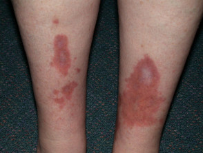
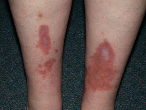
Generally, the first sign of necrobiosis lipoidica is one or more yellowish-brown plaques that appear and evolve slowly over several months, usually located on the anterior aspect of the legs (shins).
- Although the shins are the most common location (occurring bilaterally), the disorder can rarely affect other areas of the body.
- The lesions can be completely asymptomatic asymptomatic or, in some cases, may present tenderness or pain upon palpation.
- The plaques have the potential to persist persist for prolonged periods, often spanning years.
- Morphologically, they can present in round, oval shapes or have irregular contours.
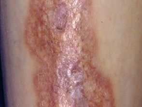
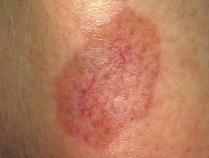
- The central area of the plaque plaque tends to become shiny, pale, and thinned, exhibiting prominent superficial blood vessels, a phenomenon known as telangiectasia. telangiectasia.
- Any minor trauma to an established plaque can lead to the development of an ulcer. These ulcers ulcers can be extremely painful or go completely unnoticed.
- Ulcers associated with necrobiosis lipoidica carry an increased risk of secondary Acute bacterial, bacterial infection, resulting in a significantly delayed healing process.
Images of Necrobiosis Lipoidica
Necrobiosis Lipoidica
Necrobiosis Lipoidica
Understanding associated factors and recognizing the early clinical characteristics of necrobiosis lipoidica is crucial for its management, especially given its strong correlation with diabetes.
Diagnosis and Confirmation of Necrobiosis Lipoidica
The diagnosis of necrobiosis lipoidica is generally established through clinical evaluation of the skin lesions. However, for definitive confirmation, a skin biopsy may be performed. biopsyPapillitis lingual transitoria cancer.
The analysis histopathological level histopathological analysis reveals characteristic findings: a granulomatous inflammatory reaction centered on the destruction of the inflammatory collagen collagen, a process termed necrobiosis or collagenolysis. It is important to note that difficulty may exist in differentiating these presentations from other pathologies such as granuloma annulare or nodule Necrobiosis lipoidica.
Rheumatoid Nodule
Treatment Options for Necrobiosis Lipoidica
- In many cases, necrobiosis lipoidica does not require active therapeutic intervention. However, when treatment is necessary to control the condition or relieve symptoms, various modalities have proven effective: topical, Application of topical steroids occlusive , frequently under plastic occlusive dressing.
- Use of intralesional steroid injections or oral administration of steroid tablets.
- Combined administration of aspirin and dipyridamole.
- Treatment with oxypentifylline tablets.
- Supplementation with niacin.
- Use of oral cyclosporine.
- Application of specific biologic agents.
- Photochemotherapy (PUVA).
- Therapy photodynamic.
- Treatment with fumaric acid esters.
When the condition progresses to ulcerative necrobiosis lipoidica, it is essential to implement a more energetic and structured treatment regimen to promote healing.


