

Merkel Cell Carcinoma: A Rare and Aggressive Form of Skin Cancer
Merkel Cell Carcinoma (MCC) represents a rare skin neoplasm, characterized by its highly aggressive potential and its tendency to metastasize to other areas of the body. Historically, this condition has been known by various names, including Toker's tumor, cutaneous neuroendocrine carcinoma, trabecular cell carcinoma, and primary small cell carcinoma of the skin.
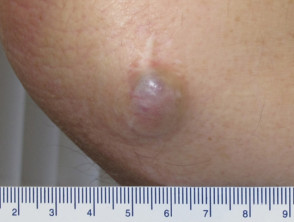
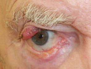
Representative Images of Merkel Cell Carcinoma
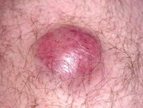
Image courtesy of Bob Rossborough
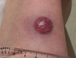
Image courtesy of Catherine Skiens
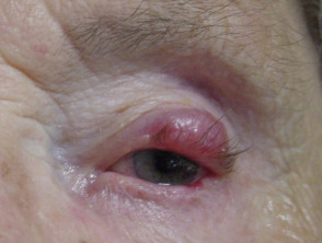
Merkel Cell Carcinoma
Epidemiological Profile: Who is most susceptible to Merkel Cell Carcinoma?
Merkel Cell Carcinoma has a low incidence, estimated at 0.23 per 100,000 people among Caucasian populations. This prevalence is considerably lower when compared to melanoma.
- Various oncology centers have reported a steady increase in the diagnosis of Merkel cell carcinomas in recent years.
- MCC predominantly affects the elderly population, with the majority of diagnoses made in individuals over 50 years of age.
- There is a slight inclination toward affecting the male sex.
- Typically, this lesion is located in areas of the body that receive frequent sun exposure, with the head and neck being primary sites of appearance.
- The risk increases drastically and the severity of the disease is greater in immunocompromised patients, including those who have received solid organ transplants, have Human Immunodeficiency Virus (HIV) infection, suffer from hematological malignancies, or are under immunosuppressive therapies such as azathioprine.
Key Clinical Features for the Diagnosis of Merkel Cell Carcinoma
Clinically, Merkel cell carcinoma usually manifests as a solitary nodule, which has a reddish tone and is characterized by its rapid growth. It is important to note that its initial presentation can be confused with more common skin cancers, such as basal cell carcinoma, although MCC notably surpasses these in proliferation speed.
These tumors have a notable capacity to spread efficiently through the lymphatic system, which can manifest as the appearance of multiple metastases in adjacent lymph nodes.
It is possible that recurrences develop around the original tumor (known as local recurrence or It is essential to remember that HNC exhibits a considerable rate of). Merkel cell carcinoma also has the capacity to spread to the lymph nodes regional lymph nodes, including those in the cervical area, the armpits and the groin. This risk increases if the primary tumors are thicker. Most of these recurrences are observed within two years following the initial diagnosis.
Essential Information about Merkel Cell Carcinoma
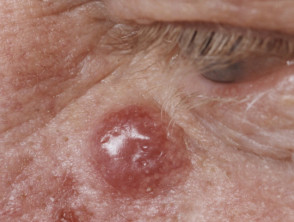
Merkel Cell Carcinoma
Understanding the aggressiveness and epidemiological profile of Merkel Cell Carcinoma is crucial for its early detection. Given its tendency for rapid metastasis, especially in immunocompromised patients, active surveillance of any rapidly growing nodule in sun-exposed areas should be a priority to optimize treatment options and improve prognosis.
What are the Causes of Merkel Cell Carcinoma?
Merkel cell polyomavirus (MCPyV) has been identified in approximately 80% of analyzed Merkel cell carcinomas. This virus is presumed to be responsible for inducing key genetic mutations mutations are being investigated. that trigger the development of the carcinoma, especially in individuals with compromised immune function.
On the other hand, those tumors classified as virus-negative have been consistently associated with high historical exposure to ultraviolet radiation, ultraviolet radiation skin , as the tumor usually manifests in skin locations that receive incidental sun exposure.
Immunosuppression represents a fundamental risk factor that enormously facilitates the development development of Merkel cell carcinomas.
Previous understandings indicated that Merkel cell carcinoma arose from the cutaneous Carcinoma Merkel cells (pressure receptor cells). However, the most recent scientific evidence suggests an origin in early B cells (lymphocyteslymphocytes). This hypothesis is based on cellular morphology morphology cellular, the expression of characteristic B cell markers in early stages, and the existence of clonal rearrangement clonal of the immunoglobulin chain. immunoglobulin.
Merkel Cell Carcinoma Diagnosis Process
Any skin lesion presenting the clinical characteristics summarized in the acronym «AEIOU,» which are identified in about 90% of patients diagnosed with this neoplasm, should prompt consideration of Merkel Cell Carcinoma:
- A: A: Asymptomatic or painless.
- E: E: Rapid expansion.
- I: Immunosuppressed (the patient).
- O: Over 50 years of age.
- U: Fair skin exposed to UV light.
The main diagnostic tool remains the biopsy biopsy pathology of the tumor, which reveals a distinctive immunohistochemistry pathology transcription of Merkel cell carcinoma. Immunohistochemistry.
is very useful: cytokeratin-20 (CK20) is positive in up to 95% of tumors, while thyroid transcription factor (TTF1) is usually negative.
It is crucial to perform a complete oncological evaluation, inspecting regional lymph nodes and employing staging studies to determine if metastatic spread has occurred.
- The recommended procedures for correct staging include:.
- Sentinel lymph node biopsy. lymph node.
Lymph node evaluation.
- To complement the evaluation, the following advanced imaging studies may be required:ultrasound.).
- Performing an ultrasound (, computed tomography (CT), magnetic resonance imaging studies. (ultrasound). Acquisition of images via X-ray, computed tomography (CT), magnetic resonance imaging (MRI) Positron Emission Tomography (PET scans.).
and Positron Emission Tomography (PET) studies.
The American Joint Committee on Cancer (AJCC) standardizes a specific system for the classification by stages (staging) of Merkel cell carcinoma.
Long-term survival is significantly more likely if the regional lymph nodes remain free of tumor cells.
What is the treatment for Merkel cell carcinoma
Treatments for Merkel Cell Carcinoma multidisciplinary. Following the diagnosis of Merkel cell carcinoma (MCC), the usual protocol involves a multidisciplinary consultation. Because 5-year survival rates for MCC are around 50%, it is crucial to implement aggressive and early treatment, which frequently combines surgery with radiotherapy.
radiotherapy. Localized
The Excision Management of Localized Disease radiotherapy Surgical excision constitutes the fundamental treatment for primary Merkel cell carcinoma. It may be necessary to remove a wide margin around the tumor; however, this technique can be omitted if supplemented with.
adjuvant therapy. lesions The primary site may be addressed with postoperative radiotherapy, especially for disease control large lesions (greater than 2 cm). Radiation treatment leads to better.
disease control both locally and regionally, optimizing long-term survival rates. prophylactically.
Relevant lymph nodes can also be removed surgically or subjected to irradiation as a
prophylactic measure. chemotherapy systemic sclerosis) Treatment if Regional Lymph Nodes are Affected.
radiotherapy. Metastatic If the cancer has spread to the lymph nodes, these can be surgically removed or treated with radiotherapy. In certain clinical scenarios, systemic
chemotherapy prognosis can also be administered to control the spread.
Management of Distant Metastatic Disease and Any Merkel cell carcinoma presenting with distant metastases is considered an extremely serious condition with a disease control, very poor prognosis. The therapeutic approach for metastatic disease focuses on improving the patient's quality of life. In specific cases, radiotherapy and/or systemic chemotherapy can be used as palliative treatment.
A positive response has been documented in up to 50% of patients with advanced Merkel cell carcinoma when treatment with immune checkpoint inhibitors disease control , specifically pembrolizumab and nivolumab, is used. The response observed to PD-1 blockade may manifest in virus-positive or negative subtypes. However, the response appears to be less significant in patients who have already received prior chemotherapy. Several clinical trials are currently underway evaluating these therapies.
It is relevant to mention that the appearance of a new Merkel cell carcinoma has been reported during treatment with a.
checkpoint inhibitor.
administered to treat other neoplasms.
In March 2017, the U.S. Food and Drug Administration (FDA) granted accelerated approval to another PD-1 / PD-L1 inhibitor agent, known as avelumab (Bavencio®), for the treatment of metastatic Merkel cell carcinoma. This approval was based on data from a clinical trial that demonstrated a response rate in one-third of treated patients.
Currently, research continues to explore experimental treatment with various innovative oncological agents.


