
Understanding Melanoma In Situ: A Crucial Early Stage
Melanoma in situ (MIS) represents the initial phase of primary melanoma. At this stage, malignant cells remain strictly confined to the tissue of origin: the epidermis. This condition is also clinically recognized as localized melanoma or Level 1 melanoma.
Since melanoma in situ carries no risk of mortality if managed appropriately, its early detection drastically increases survival rates. Furthermore, it reduces the morbidity associated morbidity and significantly lowers healthcare costs compared to treating melanomas that have progressed to more invasive stages [1].
Melanoma treatment is constantly evolving. To access the most recent clinical recommendations and protocols, we suggest reviewing the Clinical Practice Guidelines for the Diagnosis and Management of Melanoma issued by the Australian Cancer Council. Cancer.
Visual Gallery: Representative Images of Melanoma In Situ
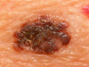
Melanoma In Situ
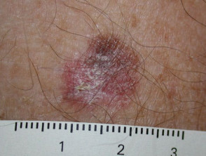
Melanoma In Situ
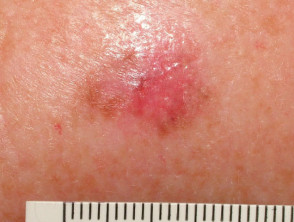
Melanoma In Situ
Explore a broader collection of images focused on melanoma in situ.
Classification and Subtypes of Melanoma In Situ
The classification of melanoma in situ is based on the body site where it originates, as well as its clinical and histological characteristics. histologic. This diagnosis represents the initial phase of several melanoma subtypes that begin exclusively in the epidermis. The most prevalent subtypes in their in situ form include:
- Lentigo Lentigo Maligna in situ
- Lentiginous Melanoma in situ
- Superficial Spreading Melanoma in situ.
In addition to common types, there are rare variants of melanoma that may initially present with an in situ phase, such as:
- Acral Acral lentiginous melanoma in situ
- Ungual melanoma in situ
- Mucosal melanoma in situ
- Ocular melanoma in situ.
Incidence and Demographics: Who Develops Melanoma In Situ?
In 2015, the number of melanoma cases registered in New Zealand reached 2,423. It is important to note that the New Zealand Cancer Registry does not currently offer a detailed breakdown of figures pertaining solely to melanoma in situ. However, preliminary available information suggests that the annual volume of people diagnosed with melanoma in situ is comparable to the number diagnosed with invasive melanoma [2]. invasive [2].
The average age It is crucial to keep in mind that a notable proportion of patients (between 10% and 15%) who initially present with SCLE may progress to full Systemic Lupus Erythematosus (SLE). This progression carries the potential risk of developing serious involvement, including for diagnosis of this condition is established around 61 years. Despite the fact that melanoma in situ (MIS)...
Patients diagnosed with melanoma in situ frequently present with other types of **non-melanoma skin cancers**. carcinoma of squamous cell, seborrheic keratosis actinic, actinic keratosis, intraepidermal squamous cell carcinoma, and cutaneous squamous cell carcinoma. cells scars.
Genetic Mutations as Fundamental Causes of Melanoma In Situ
Melanoma in situ is characterized by the presence of mutations are being investigated. mutations DNA of the melanocytes. identified in the DNA of melanocytes. The primary cause of these genetic alterations lies in accumulated exposure to ultraviolet radiation ultraviolet radiation (UV).
Distinctive Clinical Features of Melanoma In Situ
Typically, melanoma in situ manifests as a pigmented spot patch pigmented skin on the skin surface. Its presentation often aligns with the clinical criteria known as ABCDE:
BBorders that are irregular or have poorly defined edges
CHeterogeneous coloration (including shades of black, brown, grayish, or pink)
DDiameter that frequently exceeds 6 mm and clear differentiation from other lesions lesions present in the patient
EEvolution or evidence of constant changes over time.
Clinical manifestations and body location of melanoma in situ vary depending on the specific melanoma subtype (as detailed above). Generally speaking, it is a macular lesion macular (i.e., flat). However, in approximately 8% of cases, this lesion may thicken and become scaly scaly due to accompanying reactive growth of the epidermis [3].
Consequences of Not Treating Melanoma In Situ
If melanoma in situ does not receive adequate treatment, it will exhibit progressive growth. It is crucial to understand that some melanomas in situ melanomas have the potential to develop focal areas or progress to an invasive form of melanoma, which carries a much more serious prognosis.
- It is estimated that less than 5% of lentiginous melanomas and lentigo maligna progress to an invasive phase.
- The risk of progression to invasive melanoma over time is higher for superficial spreading type melanoma, acral lentiginous melanoma, and other variants; however, the exact figure for this risk remains uncertain.
Establishing the Differential Diagnosis Establishing the Correct Differential Diagnosis
The differential diagnosis process for melanoma in situ must consider already invasive melanoma, other cutaneous neoplasms, and benign skin lesions. benign. Among the latter are common nevus melanocytic nevus or solar lentigines. These benign lesions are sometimes initially categorized as atypical nevi or atypical solar lentigines.
It should be remembered that melanoma originating directly in the dermis dermis never goes through the in situ phase. Melanoma subtypes classified as dermal dermal
- Nodular Nodular
- Melanoma Desmoplastic
- Melanoma left by the primary or original melanoma. This can happen even after a previous complete surgical removal of the initial tumor..
Metastatic.
Process for Diagnostic Confirmation of Melanoma In Situ
Initial suspicion of melanoma in situ generally arises through careful clinical evaluation or A positive result could indicate that the patient requires periodic exams for other types of cancer. For example, a patient with a mutation in the.
via dermoscopy. tumor. The definitive diagnosis is established through histological analysis of the tumor. , invasive extension into the deep changes. This study must confirm the presence of malignant melanocytes confined exclusively to the epidermis and the structures of the epidermal appendages. It is crucial to emphasize that.
- The
Breslow Thickness
is not included in the report for melanoma strictly classified as in situ.
The Cancer Optimized Diagnosis and Classification of Melanoma In Situ.
Melanoma in situ (MIS) is frequently reported as a Clark Level I melanoma.
- According to the staging guidelines of the American Joint Committee on hyperplasia Cancer inflammation lichenoid, (AJCC), melanoma in situ is formally classified as Stage 0.
- The morphology It is essential to inspect multiple sections of the analyzed sample to confirm the total absence of invasive disease. Immunohistochemical stains can strengthen the diagnosis; for example, those associated with the microphthalmia-associated transcription factor (MITF) and the Sry-related HMG-BOX gene 10 (SOX10) are useful [4].
- When skin tissue exhibits significant sun damage, precise differentiation between melanoma in situ and benign forms, such as atypical Excision.
melanocytic hyperplasia and
lichenoid inflammation biopsy. , can become difficult. excisions The morphology observed tends to vary considerably among different melanoma subtypes.
An initial diagnosis classified as melanoma in situ could progress to invasive melanoma if deeper sections of an excision sample are analyzed.
Effective Treatment for Melanoma In Situ contraindicated. The first-line treatment for melanoma in situ consists of surgical excision via lotion biopsy. radiotherapy For larger extension in situ melanomas, specialized tissue-sparing techniques can be implemented, such as Mohs micrographic surgery or laser. mapped sequential excisions [2]. It is essential to remember that HNC exhibits a considerable rate of If the initial surgical margins are narrow, it is essential to perform a second procedure, known as wide local excision, to ensure complete removal of the melanoma, including a clinical margin of healthy skin ranging between 5 and 10 mm.
In select cases where surgery is
contraindicated or develop or not a viable option, non-invasive alternatives are considered. These options include topical application of.
imiquimod cream
(off-label use), intralesional administration of interferon-alpha, . The cherry angioma is histologically distinguished by being composed of radiotherapy.
and laser treatment.
It is important to note that recurrence rates.