
Maculopapular Cutaneous Mastocytosis: General Description and Histology
Cutaneous mastocytosis maculopapular cutaneous was previously known as hives pigmentosa. This is, in fact, the most frequent variant of mastocytosis observed in both adult and pediatric patients.
Unlike infantile forms, maculopapular mastocytosis in adults is unlikely to resolve spontaneously over time. There is a low, but present, risk that it may progress to systemic involvement systemic sclerosis), which implies the dissemination of the disease to multiple organ tissues.
Histological Analysis of Maculopapular Cutaneous Mastocytosis
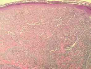
From a histological, maculopapular cutaneous mastocytosis is characterized by a marked accumulation of mast cells infiltrating mast cells located in the dermis (as illustrated in Figures 1 through 4). The study histopathological level reveals that these mast cells usually present round or fusiform morphologies.
Their microscopic characteristics include an cytoplasm abundant and strongly eosinophilic containing granules. minuscule granules, cytoplasmic borders antibodies well-defined, and nuclei. large nuclei that tend to be pale (mostly discernible in Figures 2 through 4). The concomitant presence of other inflammatory cells, such as histiocytes, eosinophils, and lymphocytes, is common. inflammatory, infections, such as and a smaller proportion of histiocytes., eosinophils y lymphocytes.
Additionally, dermal edema can be detected, especially if the dermal . The cherry angioma is histologically distinguished by being composed of lesion has undergone any type of mechanical trauma or friction.
Histopathological Illustrations of Maculopapular Cutaneous Mastocytosis
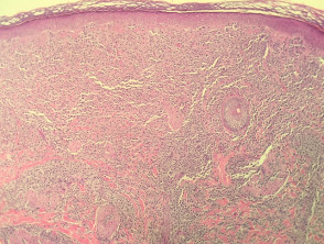
Figure 1
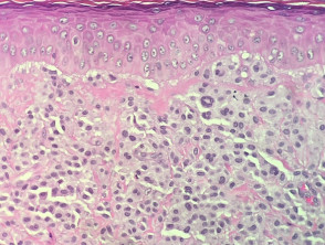
Figure 2
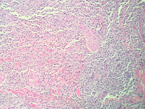
Figure 4
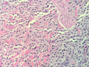
Figure 5
A detailed understanding of the histopathological features is fundamental to differentiate maculopapular cutaneous mastocytosis from other skin pathologies. The definitive diagnosis is based on the identification of these abnormal dermal mast cell clusters.
Specialized Studies for the Diagnosis of Maculopapular Cutaneous Mastocytosis
To highlight and confirm the presence of mast cells in the samples, various specialized histochemical stains are employed. Giemsa stain, toluidine blue, and tests with tryptase and Leder stain can be crucial for visualizing these cellular elements. In cases of complex diagnosis, immunohistochemistry immunohistochemistry using the CD117 marker is particularly valuable, as it offers high specificity for identifying mast cells.
Differential Diagnosis of Maculopapular Cutaneous Mastocytosis
It is essential to differentiate maculopapular cutaneous mastocytosis from other dermatological conditions that may present similar morphological or clinical characteristics. Some important diagnoses to consider include:
- Langerhans Cell Histiocytosis: This disease shares morphological and clinical presentation similarities with mastocytosis. However, immunohistochemical analysis reveals a key difference: the cells in Langerhans cell histiocytosis will be positive for CD1a, contrasting with the CD117 positivity observed in mastocytosis.
- Urticaria: Although the cutaneous infiltrate in urticaria may contain some mast cells, it is typically characterized by a much higher prevalence of neutrophils neutrophils and eosinophils, with more prominent perivascular infiltrates around blood vessels.


