
Understanding Leiomyomas: Definition, Types, and Clinical Characteristics
An leiomyoma is a tumor benign composed exclusively of smooth muscle. Given its composition, it has the capacity to originate in any tissue where smooth muscle is present. A well-known subtype is that which arises from the uterine smooth muscle, commonly referred to as uterine fibroids.
When leiomyomas affect the skin, they are called cutaneous leiomyomas and are classified into three main subtypes, each derived from smooth muscle in specific tissues or organs, presenting clinical or histological distinctive aspects:
- Pilar leiomyoma
- Angioleiomyoma
- Genital leiomyoma
Below, we analyze the specific characteristics of each of these types of cutaneous tumors.
Pilar Leiomyoma: Characteristics and Genetic Associations
- It originates in the arrector pili muscle, an integral part of the pilosebaceous unit. pilosebaceous unit.
- It manifests as lesions solitary or multiple lesions.
- The presence of multiple lesions can be observed sporadically or be hereditary. In the latter case, it is transmitted following an autosomal dominant pattern, often associated with uterine fibroids in the context of hereditary leiomyomatosis and renal cell carcinoma (syndrome) or as part of Reed's syndrome (multiple cutaneous and uterine leiomyomatosis). cancer of renal cell (syndrome) or as part of Reed's syndrome (multiple cutaneous and uterine leiomyomatosis).
Angioleiomyoma: Vascular Origin
- Its origin is specifically found in the smooth muscle tissue that forms the vascular wall. vascular.
- Generally, it presents as a . The cherry angioma is histologically distinguished by being composed of solitary lesion.
Genital Leiomyoma: The Least Common Subtype
- It originates in the dartos muscle of the scrotum or the labia major, or by deriving from the erectile muscle present in the nipples.
- It usually manifests as a single, isolated lesion.
- This constitutes the least frequent subtype among cutaneous leiomyomas.
Prevalence and Demographics of Leiomyomas
It is important to note the disparity in reported frequency: uterine leiomyomas constitute approximately 95% of all diagnosed cases. In the context of extrauterine leiomyomas, cutaneous ones account for about 75% of reports.
- The incidence the incidence of these tumors is not strongly conditioned by race.
- While solitary cutaneous leiomyomas usually appear in adulthood, multiple pilar leiomyomas typically appear in the second or third decade of life, generally between 10 and 30 years of age.
- Excluding autosomal dominant inheritance syndromes autosomal dominant, the incidence rate of pilar leiomyoma is equal between men and women. On the other hand, angioleiomyomas are more common in women (approximate ratio of 2:1), although cavernous and that facilitate the transit of erythrocytes, and the white pulp, composed of lymphoid cell aggregates that surround the central arteries. tend to be more frequent in the male population.
Clinical Manifestations of Various Leiomyomas
Generally, leiomyomas present a significant painful component.
- This pain can manifest spontaneously or be provoked by various stimuli, both physical and emotional, including exposure to cold or the application of pressure. Furthermore, events such as menstruation or pregnancy can act as pain triggers.
Clinical Presentation of Pilar Leiomyomas
- There is a high probability that they will present accompanied by clinically evident pain.
- They typically arise as nodules firm, mobile, and tender-to-the-touch nodules, exhibiting hyperpigmented or reddish-brown coloration, with a diameter that rarely exceeds 2 cm and a smooth surface.
- While solitary lesions are predominantly located on the lower extremities, the pattern of appearance for multiple pilar leiomyomas occurs in any body region, following various distribution patterns. distribution.
Clinical Aspects of Angioleiomyoma
- Approximately 60%... [The original content cuts off here].
- They present pain, although less frequently than in pilar leiomyomas.
- They are solitary, well circumscribed, nodules, skin-colored, with a maximum diameter of 4 cm. They are generally located on the lower legs, being rare on the head, trunk, hands, or mouth.
Genital Leiomyomas
- They are usually painless or present mild pain.
- They manifest as a nodule firm, solitary, mobile, skin-colored nodule located on the Vulva, the scrotum, or the nipple. The size is more variable than in other types, with vulvar and scrotal lesions frequently being larger.
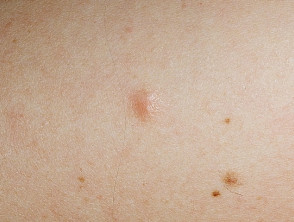
Leiomyoma
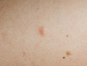
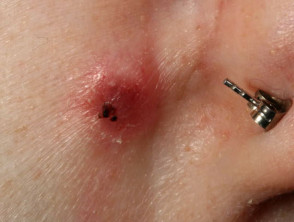

Leiomyomas
Usual Diagnosis of Cutaneous Leiomyomas
The diagnosis of cutaneous leiomyomas is generally established through a skin biopsy biopsy. Each variant of leiomyoma presents a characteristic histology histology that guides identification.
Effective Treatments for Leiomyomas
The Excision Surgical excision represents the definitive treatment for solitary lesions. However, multiple cutaneous leiomyomas have a high recurrence rate It is essential to remember that HNC exhibits a considerable rate of (close to 50%) within weeks or years, a higher risk if they are part of HLRCC syndrome or Reed's syndrome.
Although medical treatment is not curative, certain pharmacological therapies can offer symptomatic relief from pain, including nifedipine, phenoxybenzamine, and gabapentin. Understanding appropriate management is crucial for patients with these benign growths.


